Science & Chemistry
Codon Chart
Decode the standard genetic code, compare all 64 RNA codons, identify amino acids and stop signals, convert DNA coding triplets, understand anticodons, and avoid reading-frame errors.
This page uses the standard genetic code unless a table explicitly says otherwise. Translation can differ in mitochondria and some organisms, so use the sequence record’s assigned genetic-code table for annotation or research work.
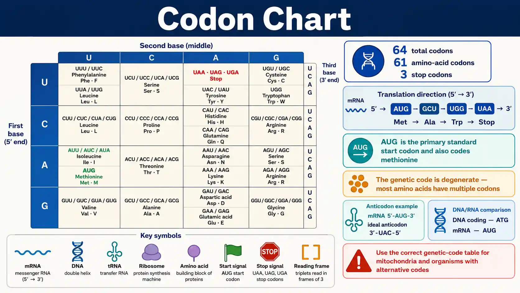
How do you read a codon chart?
Read the mRNA sequence in the 5′→3′ direction, split the correct reading frame into groups of three bases, and match each triplet to the standard genetic-code table. The standard code contains 64 triplets: 61 specify amino acids and UAA, UAG, and UGA are stop signals. See the NHGRI codon definition.
Total triplets
64 codons
Four RNA bases arranged in triplets produce 64 possible codons in the standard code table.
Sense codons
61 encode amino acids
The remaining three standard codons—UAA, UAG, and UGA—serve as translation stop signals.
Primary start
AUG → Met
AUG is the primary standard initiation codon and also encodes methionine at internal positions.
Direction rule
Read mRNA 5′→3′
Strand identity, direction, reading frame, and genetic-code table must be set before translation.
Codon answers at a glance
These direct answers cover the most common questions about triplets, reading direction, start and stop signals, degeneracy, DNA conversion, and anticodons.
What is a codon?
A codon is a three-nucleotide DNA or RNA sequence that specifies an amino acid or a translation stop signal. Codon charts are usually shown as mRNA triplets.
How many codons are in the genetic code?
There are 64 possible codons. In the standard genetic code, 61 specify amino acids and three are stop codons.
What is the standard start codon?
AUG is the primary start codon in the standard code and encodes methionine. Its start function depends on initiation context.
What are the standard stop codons?
UAA, UAG, and UGA are stop codons in NCBI translation table 1.
Which direction is mRNA read?
The ribosome reads mRNA in the 5-prime to 3-prime direction.
Why can several codons encode one amino acid?
The genetic code is degenerate, so most amino acids have more than one synonymous codon.
Which amino acids have six standard codons?
Leucine, serine, and arginine each have six codons in the standard genetic code.
Which amino acids have only one standard codon?
Methionine uses AUG and tryptophan uses UGG in the standard code.
How do DNA coding triplets relate to mRNA codons?
A DNA coding-strand triplet has the same base order as the mRNA codon after replacing DNA thymine with RNA uracil.
Is an anticodon written in the same direction as a codon?
No. Codon and anticodon pair antiparallel, so a codon written 5-prime to 3-prime pairs with its anticodon in the 3-prime to 5-prime orientation.
Does every organism use exactly the same code?
No. The standard code is nearly universal, but mitochondria and some organisms use documented alternative translation tables.
Does a codon chart show codon usage frequency?
No. A codon chart shows coding assignment; codon usage measures how often synonymous codons occur in a particular sequence set.
Standard RNA Codon Chart — All 64 Codons
The standard genetic code maps 64 mRNA triplets to 20 standard amino acids or translation termination. Codons are written 5-prime to 3-prime.
Swipe horizontally inside the table to view every column.
| mRNA codon | Assignment | 3-letter | 1-letter | Role |
|---|---|---|---|---|
| UUU | Phenylalanine | Phe | F | Amino-acid codon |
| UUC | Phenylalanine | Phe | F | Amino-acid codon |
| UUA | Leucine | Leu | L | Amino-acid codon |
| UUG | Leucine | Leu | L | Amino-acid codon |
| UCU | Serine | Ser | S | Amino-acid codon |
| UCC | Serine | Ser | S | Amino-acid codon |
| UCA | Serine | Ser | S | Amino-acid codon |
| UCG | Serine | Ser | S | Amino-acid codon |
| UAU | Tyrosine | Tyr | Y | Amino-acid codon |
| UAC | Tyrosine | Tyr | Y | Amino-acid codon |
| UAA | Stop | Ter | * | Stop signal |
| UAG | Stop | Ter | * | Stop signal |
| UGU | Cysteine | Cys | C | Amino-acid codon |
| UGC | Cysteine | Cys | C | Amino-acid codon |
| UGA | Stop | Ter | * | Stop signal |
| UGG | Tryptophan | Trp | W | Amino-acid codon |
| CUU | Leucine | Leu | L | Amino-acid codon |
| CUC | Leucine | Leu | L | Amino-acid codon |
| CUA | Leucine | Leu | L | Amino-acid codon |
| CUG | Leucine | Leu | L | Amino-acid codon |
| CCU | Proline | Pro | P | Amino-acid codon |
| CCC | Proline | Pro | P | Amino-acid codon |
| CCA | Proline | Pro | P | Amino-acid codon |
| CCG | Proline | Pro | P | Amino-acid codon |
| CAU | Histidine | His | H | Amino-acid codon |
| CAC | Histidine | His | H | Amino-acid codon |
| CAA | Glutamine | Gln | Q | Amino-acid codon |
| CAG | Glutamine | Gln | Q | Amino-acid codon |
| CGU | Arginine | Arg | R | Amino-acid codon |
| CGC | Arginine | Arg | R | Amino-acid codon |
| CGA | Arginine | Arg | R | Amino-acid codon |
| CGG | Arginine | Arg | R | Amino-acid codon |
| AUU | Isoleucine | Ile | I | Amino-acid codon |
| AUC | Isoleucine | Ile | I | Amino-acid codon |
| AUA | Isoleucine | Ile | I | Amino-acid codon |
| AUG | Methionine | Met | M | Start-capable Met codon |
| ACU | Threonine | Thr | T | Amino-acid codon |
| ACC | Threonine | Thr | T | Amino-acid codon |
| ACA | Threonine | Thr | T | Amino-acid codon |
| ACG | Threonine | Thr | T | Amino-acid codon |
| AAU | Asparagine | Asn | N | Amino-acid codon |
| AAC | Asparagine | Asn | N | Amino-acid codon |
| AAA | Lysine | Lys | K | Amino-acid codon |
| AAG | Lysine | Lys | K | Amino-acid codon |
| AGU | Serine | Ser | S | Amino-acid codon |
| AGC | Serine | Ser | S | Amino-acid codon |
| AGA | Arginine | Arg | R | Amino-acid codon |
| AGG | Arginine | Arg | R | Amino-acid codon |
| GUU | Valine | Val | V | Amino-acid codon |
| GUC | Valine | Val | V | Amino-acid codon |
| GUA | Valine | Val | V | Amino-acid codon |
| GUG | Valine | Val | V | Amino-acid codon |
| GCU | Alanine | Ala | A | Amino-acid codon |
| GCC | Alanine | Ala | A | Amino-acid codon |
| GCA | Alanine | Ala | A | Amino-acid codon |
| GCG | Alanine | Ala | A | Amino-acid codon |
| GAU | Aspartic acid | Asp | D | Amino-acid codon |
| GAC | Aspartic acid | Asp | D | Amino-acid codon |
| GAA | Glutamic acid | Glu | E | Amino-acid codon |
| GAG | Glutamic acid | Glu | E | Amino-acid codon |
| GGU | Glycine | Gly | G | Amino-acid codon |
| GGC | Glycine | Gly | G | Amino-acid codon |
| GGA | Glycine | Gly | G | Amino-acid codon |
| GGG | Glycine | Gly | G | Amino-acid codon |
RNA alphabet: A, U, C, G. Standard genetic code, NCBI translation table 1.
- • There are 64 triplets: 61 sense codons and three stop codons in the standard code.
- • AUG encodes methionine and is the primary initiation codon, but internal AUG codons also encode methionine.
Download or export
Amino Acids Grouped by Their Standard Codons
The genetic code is degenerate: most amino acids are encoded by more than one codon, while methionine and tryptophan each have one standard codon.
Swipe horizontally inside the table to view every column.
| Assignment | 3-letter | 1-letter | Standard RNA codons | Count |
|---|---|---|---|---|
| Phenylalanine | Phe | F | UUU, UUC | 2 |
| Leucine | Leu | L | UUA, UUG, CUU, CUC, CUA, CUG | 6 |
| Isoleucine | Ile | I | AUU, AUC, AUA | 3 |
| Methionine | Met | M | AUG | 1 |
| Valine | Val | V | GUU, GUC, GUA, GUG | 4 |
| Serine | Ser | S | UCU, UCC, UCA, UCG, AGU, AGC | 6 |
| Proline | Pro | P | CCU, CCC, CCA, CCG | 4 |
| Threonine | Thr | T | ACU, ACC, ACA, ACG | 4 |
| Alanine | Ala | A | GCU, GCC, GCA, GCG | 4 |
| Tyrosine | Tyr | Y | UAU, UAC | 2 |
| Histidine | His | H | CAU, CAC | 2 |
| Glutamine | Gln | Q | CAA, CAG | 2 |
| Asparagine | Asn | N | AAU, AAC | 2 |
| Lysine | Lys | K | AAA, AAG | 2 |
| Aspartic acid | Asp | D | GAU, GAC | 2 |
| Glutamic acid | Glu | E | GAA, GAG | 2 |
| Cysteine | Cys | C | UGU, UGC | 2 |
| Tryptophan | Trp | W | UGG | 1 |
| Arginine | Arg | R | CGU, CGC, CGA, CGG, AGA, AGG | 6 |
| Glycine | Gly | G | GGU, GGC, GGA, GGG | 4 |
| Stop | Ter | * | UAA, UAG, UGA | 3 |
Counts refer to NCBI standard genetic code assignments.
- • Leucine, serine, and arginine each have six standard codons.
- • Degeneracy does not mean a codon is ambiguous: each standard codon has one standard assignment in a specified genetic code table.
Download or export
Why the code is called degenerate
Most amino acids have more than one synonymous codon. Leucine, serine, and arginine each have six standard codons, while methionine and tryptophan each have only one. Degeneracy gives multiple triplets the same amino-acid assignment; it does not make an individual codon uncertain within a specified genetic-code table.
Wobble in codon-anticodon recognition helps some tRNAs read more than one synonymous codon, especially through flexibility at the third codon position.
Start and Stop Codon Chart
Initiation and termination are context-dependent translation signals. The standard code has three stop codons and uses AUG as the primary initiation codon.
Swipe horizontally inside the table to view every column.
| Codon | Standard assignment | Translation role | Important note |
|---|---|---|---|
| AUG | Methionine (Met, M) | Primary start codon and internal methionine codon | Start role depends on initiation context; internal AUG codes Met |
| UAA | Stop | Termination signal | No standard amino acid assignment in translation table 1 |
| UAG | Stop | Termination signal | No standard amino acid assignment in translation table 1 |
| UGA | Stop | Termination signal | Stop in the standard code; Trp in several mitochondrial and microbial codes |
mRNA codons, written 5-prime to 3-prime.
- • AUG is not exclusively a start signal; it also encodes methionine within coding sequences.
- • Alternative initiation codons exist in some organisms and organelles, so sequence context and the assigned translation table matter.
Download or export
Codon Family and Degeneracy Patterns
Many synonymous codons share their first two bases and vary at the third position. IUPAC-style ambiguity letters are used here only as shorthand for grouped codons.
Swipe horizontally inside the table to view every column.
| Pattern | Assignment | Pattern meaning | Expanded codons | Interpretation |
|---|---|---|---|---|
| UUY | Phe | Y = U or C | UUU, UUC | Two-codon family |
| UUR | Leu | R = A or G | UUA, UUG | Two-codon family |
| UCN | Ser | N = any base | UCU, UCC, UCA, UCG | Four-codon family |
| UAY | Tyr | Y = U or C | UAU, UAC | Two-codon family |
| UAR | Stop | R = A or G | UAA, UAG | Two stop codons |
| UGY | Cys | Y = U or C | UGU, UGC | Two-codon family |
| CUN | Leu | N = any base | CUU, CUC, CUA, CUG | Four-codon family |
| CCN | Pro | N = any base | CCU, CCC, CCA, CCG | Four-codon family |
| CGN | Arg | N = any base | CGU, CGC, CGA, CGG | Four-codon family |
| ACN | Thr | N = any base | ACU, ACC, ACA, ACG | Four-codon family |
| GUN | Val | N = any base | GUU, GUC, GUA, GUG | Four-codon family |
| GCN | Ala | N = any base | GCU, GCC, GCA, GCG | Four-codon family |
| GGN | Gly | N = any base | GGU, GGC, GGA, GGG | Four-codon family |
| AGR | Arg | R = A or G | AGA, AGG | Adds to CGN to give six Arg codons |
Y = pyrimidine (U/C); R = purine (A/G); N = any standard RNA base.
- • The third codon position often tolerates synonymous changes, but not every third-position change is synonymous.
- • Serine, leucine, and arginine use codons from more than one first-two-base family.
Download or export
Codon lookup and strand converter
Enter one mRNA codon or one DNA coding-strand triplet. The lookup uses the standard genetic code and shows the corresponding strand notation.
DNA Coding Strand, Template Strand and mRNA Examples
The DNA coding strand matches the mRNA base order except that DNA uses T instead of U. The template strand is complementary and antiparallel to the mRNA.
Swipe horizontally inside the table to view every column.
| mRNA codon 5′→3′ | DNA coding 5′→3′ | DNA template 3′→5′ | Standard assignment |
|---|---|---|---|
| AUG | ATG | TAC | Methionine |
| UUU | TTT | AAA | Phenylalanine |
| UUA | TTA | AAT | Leucine |
| UCG | TCG | AGC | Serine |
| UAU | TAT | ATA | Tyrosine |
| UGG | TGG | ACC | Tryptophan |
| CUA | CTA | GAT | Leucine |
| AUA | ATA | TAT | Isoleucine |
| AAA | AAA | TTT | Lysine |
| AGG | AGG | TCC | Arginine |
| GCU | GCT | CGA | Alanine |
| UGA | TGA | ACT | Stop |
Sequences are shown in explicit strand directions.
- • Do not simply replace T with U when starting from a DNA template strand; first account for complementarity and direction.
- • GenBank genetic-code tables often display DNA triplets with T by historical convention.
Download or export
DNA coding strand versus DNA template strand
A DNA coding strand has the same sequence order as the mRNA except DNA uses T where RNA uses U. A DNA template strand is complementary and antiparallel to the mRNA, so template input must be complemented and oriented correctly before using an mRNA codon chart.
For example, DNA coding 5′-ATG-3′ corresponds to mRNA 5′-AUG-3′, while the paired DNA template is 3′-TAC-5′.
Codon and Ideal Complementary Anticodon Examples
Codon and anticodon strands pair antiparallel. This table shows the ideal Watson-Crick complement, not every biologically used tRNA anticodon.
Swipe horizontally inside the table to view every column.
| mRNA codon | Ideal complement on anticodon | Amino acid | Interpretation |
|---|---|---|---|
| 5′-AUG-3′ | 3′-UAC-5′ | Methionine | Ideal complementary pairing; real tRNAs may use wobble or modified bases |
| 5′-UUU-3′ | 3′-AAA-5′ | Phenylalanine | Ideal complementary pairing; real tRNAs may use wobble or modified bases |
| 5′-UUC-3′ | 3′-AAG-5′ | Phenylalanine | Ideal complementary pairing; real tRNAs may use wobble or modified bases |
| 5′-GCU-3′ | 3′-CGA-5′ | Alanine | Ideal complementary pairing; real tRNAs may use wobble or modified bases |
| 5′-GAA-3′ | 3′-CUU-5′ | Glutamic acid | Ideal complementary pairing; real tRNAs may use wobble or modified bases |
| 5′-AAA-3′ | 3′-UUU-5′ | Lysine | Ideal complementary pairing; real tRNAs may use wobble or modified bases |
| 5′-UGG-3′ | 3′-ACC-5′ | Tryptophan | Ideal complementary pairing; real tRNAs may use wobble or modified bases |
| 5′-CAU-3′ | 3′-GUA-5′ | Histidine | Ideal complementary pairing; real tRNAs may use wobble or modified bases |
| 5′-ACG-3′ | 3′-UGC-5′ | Threonine | Ideal complementary pairing; real tRNAs may use wobble or modified bases |
| 5′-CGA-3′ | 3′-GCU-5′ | Arginine | Ideal complementary pairing; real tRNAs may use wobble or modified bases |
| 5′-GUU-3′ | 3′-CAA-5′ | Valine | Ideal complementary pairing; real tRNAs may use wobble or modified bases |
| 5′-UAC-3′ | 3′-AUG-5′ | Tyrosine | Ideal complementary pairing; real tRNAs may use wobble or modified bases |
mRNA codon is 5′→3′; paired anticodon complement is shown 3′→5′.
- • Real tRNA decoding can use wobble pairing and modified bases, especially at the third codon position.
- • A stop codon is recognized by release factors rather than a standard aminoacyl-tRNA anticodon.
Download or export
Why an anticodon is not always a simple one-to-one lookup
Codon and anticodon strands pair antiparallel, but real tRNA recognition can use wobble pairing and modified nucleotides. One tRNA can therefore recognize more than one synonymous codon. Review NCBI’s translation overview.
How Sequence Changes Affect Codons and Translation
A nucleotide change can be synonymous, missense, nonsense, stop-loss, or frame-altering depending on its position and effect on the reading frame.
Swipe horizontally inside the table to view every column.
| Change type | What changes | Simple example | Translation result | Key point |
|---|---|---|---|---|
| Synonymous substitution | Codon changes but amino acid stays the same | GAA → GAG | Glu → Glu | Often possible because the code is degenerate |
| Missense substitution | Codon changes to a different amino acid | GAA → GUA | Glu → Val | Protein effect depends on residue role and substitution |
| Nonsense substitution | Sense codon changes to a stop codon | UAU → UAA | Tyr → Stop | Can truncate translation |
| Stop-loss substitution | Stop codon changes to a sense codon | UAG → CAG | Stop → Gln | Can extend translation until a later stop |
| Start-codon change | AUG changes to another triplet | AUG → ACG | Initiation signal altered | Effect depends on transcript context and alternative initiation |
| Frameshift insertion | Nucleotides inserted in a number not divisible by 3 | +1 nucleotide | Reading frame changes | All downstream codons can change |
| Frameshift deletion | Nucleotides deleted in a number not divisible by 3 | −2 nucleotides | Reading frame changes | All downstream codons can change |
| In-frame insertion/deletion | Adds or removes a multiple of 3 nucleotides | +3 or −3 nucleotides | Adds/removes residue(s) | Reading frame is preserved |
Examples use mRNA notation and illustrate principles, not clinical interpretation.
- • A codon chart predicts the direct coding assignment, not the biological severity of a variant.
- • Frameshifts arise when coding insertions or deletions are not multiples of three nucleotides.
Download or export
Selected Genetic Code Variants
The standard code is nearly universal, but mitochondria and some organisms use documented alternative assignments. Always use the correct NCBI translation table for the sequence source.
Swipe horizontally inside the table to view every column.
| Genetic code | AUA | UGA | Other notable difference | Use context |
|---|---|---|---|---|
| Standard code (NCBI 1) | AUA = Ile | UGA = Stop | AGA/AGG = Arg | Primary reference for nuclear coding in most organisms |
| Vertebrate mitochondrial (NCBI 2) | AUA = Met | UGA = Trp | AGA/AGG = Stop | Mitochondrial coding differs at several triplets |
| Yeast mitochondrial (NCBI 3) | AUA = Met | UGA = Trp | CUN = Thr | Mitochondrial code in specified yeasts; not the standard nuclear code |
| Mold/protozoan/coelenterate mitochondrial + Mycoplasma/Spiroplasma (NCBI 4) | AUA = Ile | UGA = Trp | AGA/AGG = Arg | Shows why organism and genetic-code table must be known |
Representative differences only; see the NCBI Genetic Codes resource for the complete current list.
- • Do not apply vertebrate mitochondrial assignments to nuclear genes.
- • NCBI tracks multiple nuclear, mitochondrial, plastid, and other organelle code tables.
Download or export
The standard code is not the only genetic code
Mitochondria and some organisms use documented alternative assignments. In vertebrate mitochondrial translation table 2, UGA is assigned tryptophan, AUA methionine, and AGA/AGG termination. NCBI also notes that actual mitochondrial termination mechanisms can be more nuanced, so sequence annotation should follow the assigned table and organism context. Check the NCBI genetic-code tables.
Codon Translation Workflow and Reading-Frame Checklist
Use this sequence to avoid strand, direction, and reading-frame errors before translating a nucleotide sequence.
Swipe horizontally inside the table to view every column.
| Step | Question | Action | Why it matters |
|---|---|---|---|
| 1 | Identify the sequence type | mRNA, DNA coding strand, or DNA template strand | A codon chart is normally written for mRNA triplets |
| 2 | Set direction | Read mRNA 5′→3′ | Triplet order changes if direction is reversed |
| 3 | Choose the reading frame | Start at the biologically defined initiation position | A one-base shift changes every downstream triplet |
| 4 | Split into triplets | Group bases three at a time | Codons are nonoverlapping in a given reading frame |
| 5 | Convert DNA coding strand if needed | Replace T with U | Coding DNA and mRNA have the same base order except T/U |
| 6 | Decode each codon | Use one standard code table consistently | Do not mix mitochondrial and standard assignments |
| 7 | Mark start and stop context | AUG is the primary standard start; UAA/UAG/UGA are standard stops | AUG can also occur internally as methionine |
| 8 | Report amino-acid symbols consistently | Use full name, three-letter, or one-letter notation as requested | IUPAC-IUB symbols standardize sequence notation |
Workflow for standard-code educational translation.
- • A sequence can have multiple possible reading frames, but biological translation uses the frame established by transcript and initiation context.
- • A codon chart alone cannot identify which open reading frame is biologically expressed.
Download or export
Common Codon Chart Mistakes and Corrections
Most codon-chart mistakes come from strand direction, DNA-versus-RNA notation, reading-frame shifts, or using the wrong genetic code.
Swipe horizontally inside the table to view every column.
| Mistake | Why it fails | Better approach | Example |
|---|---|---|---|
| Reading mRNA 3′→5′ | Translation proceeds along mRNA in the 5′→3′ direction | Write the sequence direction before grouping codons | 5′-AUG-GCU-3′ gives AUG then GCU |
| Using a DNA template triplet as if it were mRNA | The template is complementary and antiparallel | Convert the template to mRNA first | 3′-TAC-5′ corresponds to 5′-AUG-3′ |
| Replacing U with T in an mRNA chart without labeling the strand | DNA coding and template strands are different | State “DNA coding strand” when using T-based codons | ATG coding DNA corresponds to AUG mRNA |
| Treating AUG as only a start signal | AUG also codes methionine within the open reading frame | Separate codon assignment from initiation context | Internal AUG = Met |
| Giving a tRNA anticodon for a stop codon | Standard termination uses release factors, not an aminoacyl-tRNA | Label UAA, UAG and UGA as stop signals | UGA has no standard amino-acid tRNA in table 1 |
| Assuming every organism uses table 1 | Alternative codes exist | Check the organism, organelle and translation-table assignment | Vertebrate mitochondria translate UGA as Trp |
| Ignoring the reading frame | Changing the starting base changes every triplet | Group codons from the biologically relevant frame | AUGGCU differs from UGG… if shifted one base |
| Assuming synonymous codons are used equally | Codon usage frequency varies among genes and organisms | Separate genetic-code assignment from codon-usage frequency | GCU/GCC/GCA/GCG all encode Ala but may differ in usage |
Use explicit strand direction, sequence type, reading frame, and translation table.
- • Codon assignment and codon usage are different concepts.
- • For research or annotation work, follow the sequence record’s translation-table metadata.
Download or export
Codon assignment is not the same as codon usage
The genetic code answers what a triplet means. Codon usage answers how frequently synonymous codons appear in a particular genome, gene set, tissue, expression system, or organism. A standard codon chart therefore cannot tell you which synonymous codon is most frequent without a separate codon-usage dataset.
Frequently asked questions
What is a codon?
A codon is a sequence of three nucleotides in DNA or RNA that specifies an amino acid or a translation stop signal. Codon charts are usually written as mRNA triplets.
How many codons are there?
There are 64 possible three-base codons because four standard RNA bases can occupy each of three positions. In the standard code, 61 specify amino acids and three are stop codons.
What are the three stop codons?
UAA, UAG, and UGA are stop codons in the standard genetic code. They signal translation termination rather than a standard amino acid.
What is the start codon?
AUG is the primary start codon in the standard code and encodes methionine. Its start role depends on initiation context, because internal AUG codons also encode methionine.
Why do several codons encode the same amino acid?
The genetic code is degenerate, meaning most amino acids have multiple synonymous codons. This redundancy is especially common at the third codon position.
Which amino acids have six codons?
Leucine, serine, and arginine each have six codons in the standard genetic code.
Which amino acids have only one codon?
Methionine is encoded by AUG and tryptophan by UGG in the standard genetic code.
Do DNA and RNA codon charts use the same letters?
The assignments are equivalent for a DNA coding strand after replacing T with U. RNA uses uracil, while DNA uses thymine.
How do I convert a DNA template strand to mRNA?
Build the complementary RNA sequence antiparallel to the DNA template, then write the mRNA 5-prime to 3-prime before splitting it into codons.
What is an anticodon?
An anticodon is a three-nucleotide sequence in tRNA that base-pairs antiparallel with an mRNA codon. Wobble and modified bases mean actual tRNA recognition can be more flexible than a simple complement table.
What does wobble mean in the genetic code?
Wobble is relaxed pairing between the third base of the codon and the first base of the tRNA anticodon, allowing some tRNAs to recognize more than one synonymous codon.
Is the genetic code universal?
The genetic code is nearly universal, but documented alternative codes occur in mitochondria and some organisms. Use the correct translation table for the sequence source.
What is a synonymous codon change?
A synonymous change alters a codon without changing the encoded amino acid in the chosen genetic code.
What is a nonsense codon change?
A nonsense change converts an amino-acid codon into a stop codon, which can terminate translation earlier.
Why does reading frame matter?
Codons are read as consecutive nonoverlapping triplets. Starting one nucleotide earlier or later creates a different set of codons and usually a different translation.
Does a codon chart show codon usage frequency?
No. A codon chart shows assignment, while codon usage describes how often synonymous codons occur in a particular gene or organism.
Related ChartsLoom references
Use the Amino Acid Chart to compare amino-acid names, sequence symbols, and side-chain properties. The Atomic Radius Chart provides a complementary periodic-property reference for chemistry study.
Sources
National Human Genome Research Institute — Codon — Genetics Glossary
Defines a codon as a three-nucleotide DNA or RNA sequence and states that 64 codons exist, with 61 specifying amino acids and three serving as stop signals.
https://www.genome.gov/genetics-glossary/Codon
National Center for Biotechnology Information — The Genetic Codes — Standard Code
Provides NCBI translation table 1, including standard DNA-triplet assignments, AUG initiation, and documented alternative genetic codes.
https://www.ncbi.nlm.nih.gov/Taxonomy/Utils/wprintgc.cgi?chapter=tgencodes
NCBI Bookshelf — Expression of Genetic Information — The Genetic Code
Explains how all 64 triplets were assigned, including 61 amino-acid codons, three stop codons, degeneracy, and near universality.
https://www.ncbi.nlm.nih.gov/books/NBK9842/
NCBI Bookshelf — Translation of mRNA
Explains tRNA anticodon pairing, wobble at the third codon position, and how redundancy relates to codon recognition.
https://www.ncbi.nlm.nih.gov/books/NBK9849/
NCBI Bookshelf — From RNA to Protein
Explains that codons are read as consecutive triplets in one reading frame and that mRNA is translated in the 5-prime to 3-prime direction.
https://www.ncbi.nlm.nih.gov/books/NBK26829/
IUPAC-IUB Joint Commission on Biochemical Nomenclature — Amino-Acid One-Letter and Three-Letter Symbols
Provides standardized amino-acid sequence symbols used alongside codon assignments.
https://iupac.qmul.ac.uk/AminoAcid/A2021.html
IUPAC Nomenclature — Nucleic Acid Symbols
Provides conventions for representing nucleotide sequences and their directionality.
https://iupac.qmul.ac.uk/misc/naabb.html