Biology and human anatomy reference
Muscle Groups Chart
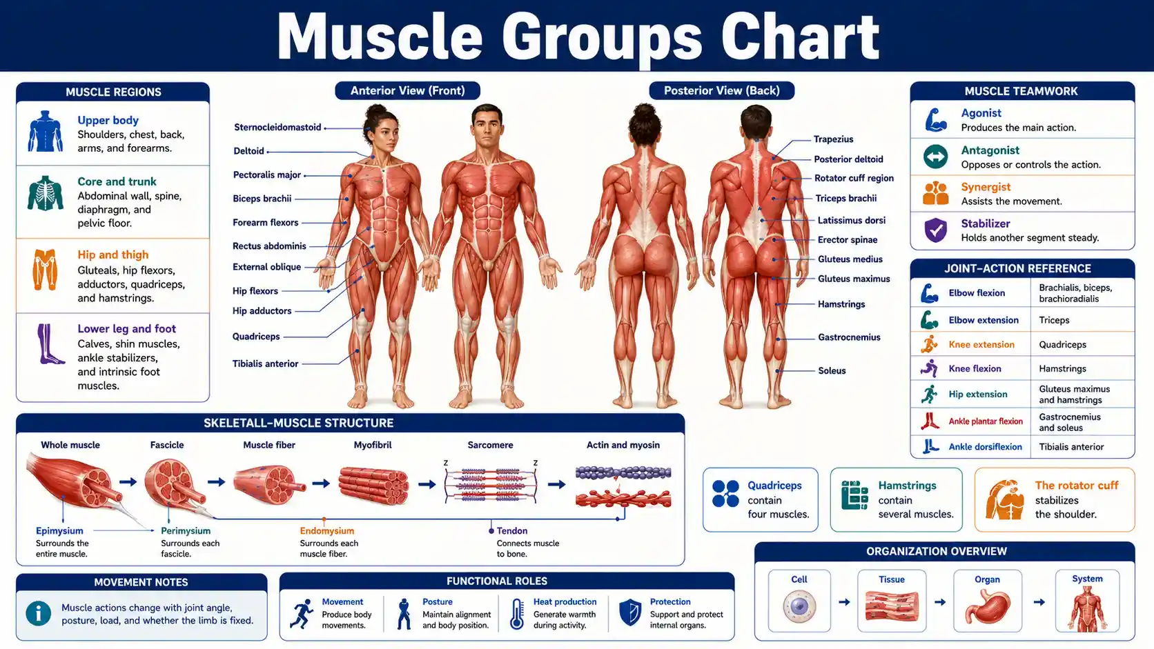
This muscle groups chart identifies major skeletal muscles by region, explains the joint actions they produce, and shows how movers, antagonists, synergists, and stabilizers coordinate human movement.
Muscle actions depend on posture, joint angle, load, and whether a limb is fixed. This anatomy reference cannot identify the cause of pain, weakness, numbness, swelling, or loss of movement.
What are the major muscle groups?
Major practical groups include the neck, shoulders, chest, back, arms, forearms, abdominal wall, spinal muscles, gluteals, hip muscles, quadriceps, hamstrings, calves, and the muscles of the lower leg and foot. Anatomists also organize skeletal muscles as axial or appendicular and by body compartment, attachments, fiber direction, and joint action.
Main tissue focus
Skeletal muscle
Muscle-group charts usually map voluntary skeletal muscles that move and stabilize the skeleton.
Organization
Axial and appendicular
Axial muscles act mainly on the head and trunk, while appendicular muscles position and move the limbs and girdles.
Movement rule
Muscles work in teams
Prime movers, antagonists, synergists, and stabilizers coordinate nearly every purposeful movement.
Counting limit
No fixed group total
Muscle groups change with the anatomical, regional, functional, clinical, or training classification being used.
Essential muscle-group questions
These concise answers establish the main anatomical definitions and limits needed to use the detailed muscle charts correctly.
What is a muscle group?
A muscle group is a practical collection of muscles that share a body region, compartment, joint action, or functional task.
How many skeletal muscles are in the body?
No universal count exists because sources handle small, variable, paired, and multi-bellied muscles differently; educational estimates commonly exceed 600.
What are the largest practical muscle groups?
Common large groups include the gluteals, quadriceps, hamstrings, back muscles, chest muscles, and calf complex, but size depends on how groups are defined.
Are quadriceps one muscle?
No. The quadriceps contain rectus femoris, vastus lateralis, vastus medialis, and vastus intermedius.
Are hamstrings one muscle?
No. The hamstrings generally include biceps femoris, semitendinosus, and semimembranosus.
What muscles form the rotator cuff?
Supraspinatus, infraspinatus, teres minor, and subscapularis form the rotator cuff.
What is the main muscle of quiet breathing?
The diaphragm is the primary muscle of quiet inspiration, assisted by rib-cage and accessory muscles when demand increases.
What muscles stabilize the pelvis during walking?
Gluteus medius and gluteus minimus are major pelvic stabilizers during single-leg support, with help from trunk and hip muscles.
What is an agonist?
An agonist produces a major share of a chosen action, but the identity of the agonist can change with joint angle, posture, and load.
What is an antagonist?
An antagonist produces or controls the opposite action and often remains active to slow movement or improve joint stability.
Does soreness prove muscle growth?
No. Soreness does not directly measure muscle growth, training quality, or recovery.
Can a muscle chart diagnose pain?
No. Pain may arise from muscle, tendon, joint, ligament, bone, nerve, fascia, blood vessel, or a referred source.
Major Muscle Groups, Locations, and Primary Actions
This overview groups commonly studied skeletal muscles by body region. Each listed group contains multiple muscles and usually performs more than one action.
Swipe horizontally inside the table to view every column.
| Muscle group | Main muscles | Body region | Primary actions | Common movement example |
|---|---|---|---|---|
| Neck flexors and extensors | Sternocleidomastoid, scalenes, splenius, upper trapezius | Anterior and posterior neck | Flex, extend, rotate, and laterally flex the head and neck | Looking down, up, or over a shoulder |
| Trapezius and scapular stabilizers | Trapezius, rhomboids, levator scapulae, serratus anterior | Upper back and shoulder girdle | Elevate, depress, retract, protract, and upwardly rotate the scapula | Reaching overhead or drawing the shoulder blades together |
| Deltoids | Anterior, middle, and posterior deltoid fibers | Shoulder cap | Flex, abduct, extend, and rotate the arm depending on fiber region | Raising the arm to the front, side, or rear |
| Chest muscles | Pectoralis major and pectoralis minor | Anterior thorax | Adduct and internally rotate the arm; position the scapula | Pushing an object away |
| Back pulling muscles | Latissimus dorsi, teres major, posterior deltoid | Posterior trunk and shoulder | Extend, adduct, and internally rotate the arm | Pulling the arms downward or backward |
| Rotator cuff | Supraspinatus, infraspinatus, teres minor, subscapularis | Deep shoulder | Stabilize the humeral head and assist abduction and rotation — Stability is a primary role | Controlling the shoulder during reaching |
| Elbow flexors | Biceps brachii, brachialis, brachioradialis | Anterior arm and lateral forearm | Flex the elbow; biceps also supinates the forearm | Bringing the hand toward the shoulder |
| Elbow extensors | Triceps brachii and anconeus | Posterior arm | Extend the elbow; long head also assists shoulder extension | Straightening the elbow during a push |
| Forearm and hand muscles | Wrist and finger flexors, extensors, pronators, supinators, intrinsic hand muscles | Forearm and hand | Move and stabilize the wrist, hand, fingers, and thumb | Gripping, typing, or turning the palm |
| Abdominal wall | Rectus abdominis, external oblique, internal oblique, transversus abdominis | Anterior and lateral trunk | Flex and rotate the trunk, compress the abdomen, and manage pressure | Curling the trunk or bracing during lifting |
| Spinal extensors | Erector spinae, multifidus, semispinalis | Deep and superficial posterior trunk | Extend, laterally flex, rotate, and stabilize the vertebral column | Maintaining an upright posture |
| Respiratory muscles | Diaphragm, intercostals, accessory breathing muscles | Thorax and neck | Change thoracic volume to move air — Includes the main breathing muscle | Quiet or forceful breathing |
| Gluteal muscles | Gluteus maximus, medius, and minimus | Posterior and lateral hip | Extend, abduct, and rotate the hip; stabilize the pelvis | Rising from a chair or balancing on one leg |
| Hip flexors | Iliopsoas, rectus femoris, sartorius, tensor fasciae latae | Anterior hip and thigh | Flex the hip; some members also rotate or abduct the thigh | Lifting the knee |
| Hip adductors | Adductor longus, brevis, magnus, gracilis, pectineus | Medial thigh | Adduct the thigh and assist hip stabilization | Drawing the leg toward the midline |
| Quadriceps | Rectus femoris, vastus lateralis, vastus medialis, vastus intermedius | Anterior thigh | Extend the knee; rectus femoris also flexes the hip — Four-muscle knee extensor group | Standing up or straightening the knee |
| Hamstrings | Biceps femoris, semitendinosus, semimembranosus | Posterior thigh | Flex the knee and extend the hip | Pulling the heel backward or accelerating in a run |
| Calf plantar flexors | Gastrocnemius and soleus | Posterior lower leg | Plantar flex the ankle; gastrocnemius also assists knee flexion | Rising onto the toes |
| Anterior lower-leg muscles | Tibialis anterior and toe extensors | Anterior lower leg | Dorsiflex the ankle and extend the toes; tibialis anterior also inverts the foot | Lifting the forefoot during walking |
| Lateral and deep lower-leg muscles | Fibularis muscles, tibialis posterior, toe flexors | Lateral and posterior lower leg | Evert or invert the foot, plantar flex the ankle, and support foot arches | Stabilizing the foot on uneven ground |
Actions describe common open-chain movements from standard anatomical position. A muscle may act differently when the limb is fixed or when several joints move together.
- • A muscle group is a practical regional or functional category, not a single anatomical structure.
- • Most movements require coordinated activity from several prime movers, synergists, antagonists, and stabilizers.
- • Surface diagrams emphasize visible muscles and cannot show every deep muscle or three-dimensional attachment.
Download or export
Functional anatomy map
Muscle groups link body regions instead of working in isolation
A useful muscle chart combines region, depth, attachment, joint action, nerve supply, and task. Surface anatomy shows only part of the system because many important stabilizers lie beneath larger muscles.
Upper body
Scapula, shoulder, arm, forearm, and hand
Upper-limb motion depends on the scapular stabilizers, rotator cuff, deltoids, chest and back muscles, elbow groups, and wrist and hand muscles working as linked teams.
Core and trunk
Breathing, pressure, posture, and load transfer
The diaphragm, abdominal wall, pelvic floor, spinal muscles, and hip stabilizers coordinate motion and resist unwanted movement.
Hip and thigh
Pelvic control and powerful limb movement
Gluteals, hip flexors, adductors, quadriceps, and hamstrings move the hip and knee while stabilizing the pelvis during standing and gait.
Lower leg and foot
Ankle control, toe clearance, arches, and push-off
Anterior, lateral, and posterior leg compartments work with intrinsic foot muscles to adapt to the surface and move the body forward.
Movement
Prime movers create a major share of an action, but their contribution changes with joint position and task.
Control
Antagonists and eccentric controllers slow motion, absorb load, and prevent a segment from moving too far or too quickly.
Stability
Fixators and stabilizers create a steady base so another joint or body segment can move efficiently.
Muscle names do not define one permanent movement role
The OpenStax muscle-interaction reference explains that origins, insertions, joint position, agonists, antagonists, synergists, fixators, and fascicle arrangement all influence movement. A muscle can create motion in one task and stabilize or decelerate the same joint in another.
Anterior and Posterior Surface Muscle Landmarks
Front and back views help identify superficial muscle groups. Deep muscles may be hidden by larger surface muscles and require cross-sectional or layered diagrams.
Swipe horizontally inside the table to view every column.
| View | Region | Prominent superficial muscles | Landmark clue | Important deeper structure |
|---|---|---|---|---|
| Anterior | Neck | Sternocleidomastoid | Oblique band from sternum and clavicle toward the mastoid region | Scalenes |
| Anterior | Shoulder and chest | Deltoid and pectoralis major | Rounded shoulder cap and broad fan over the upper chest | Pectoralis minor and rotator-cuff tendons |
| Anterior | Arm | Biceps brachii | Prominent anterior upper-arm belly during elbow flexion | Brachialis — Deep primary elbow flexor |
| Anterior | Abdomen | Rectus abdominis and external oblique | Vertical midline columns and lateral diagonal fibers | Internal oblique and transversus abdominis |
| Anterior | Hip and thigh | Tensor fasciae latae, sartorius, quadriceps, adductors | Long sartorius crosses the thigh diagonally; quadriceps dominate the front | Iliopsoas and deep hip rotators |
| Anterior | Lower leg | Tibialis anterior and toe extensors | Tibialis anterior lies just lateral to the tibial crest | Deep posterior flexors |
| Posterior | Upper back | Trapezius, posterior deltoid, latissimus dorsi | Trapezius spans neck to mid-back; latissimus forms the posterior axillary fold | Rhomboids and rotator cuff — Deep scapular and shoulder stabilizers |
| Posterior | Arm | Triceps brachii | Large posterior upper-arm group converging on the elbow | Radial-nerve and deep fascial relationships |
| Posterior | Spine | Erector spinae columns | Long vertical muscle masses on both sides of the vertebral column | Multifidus and smaller transversospinal muscles |
| Posterior | Hip | Gluteus maximus and gluteus medius | Gluteus maximus forms most posterior contour; medius appears superior-laterally | Gluteus minimus and short external rotators |
| Posterior | Thigh | Hamstrings | Long muscles between the gluteal region and back of the knee | Adductor magnus |
| Posterior | Lower leg | Gastrocnemius and soleus | Gastrocnemius creates the upper calf contour; soleus extends lower and deeper | Tibialis posterior and toe flexors |
Surface visibility changes with body composition, position, contraction, lighting, and individual anatomy. Visible contour does not define the entire muscle boundary.
- • Anterior and posterior labels describe the standard anatomical position, not every exercise posture.
- • Some large muscles cover smaller stabilizers that remain essential for joint control.
- • Palpation and movement testing require anatomical training and cannot diagnose injury by themselves.
Download or export
Upper-Body Muscle Groups by Joint and Movement
Upper-body movement depends on coordination between the scapula, shoulder, elbow, forearm, wrist, and hand rather than isolated contraction of one visible muscle.
Swipe horizontally inside the table to view every column.
| Region or group | Key muscles | Main joint actions | Stabilization role | Typical functional task |
|---|---|---|---|---|
| Scapular elevators and retractors | Upper and middle trapezius, rhomboids, levator scapulae | Elevate and retract the scapula | Hold the scapula against the thorax and position the shoulder socket | Carrying, rowing, and postural control |
| Scapular protractors and upward rotators | Serratus anterior with upper and lower trapezius | Protract and upwardly rotate the scapula | Maintain scapular contact with the rib cage during arm elevation — Scapular stability during elevation | Reaching or pressing overhead |
| Shoulder abductors | Middle deltoid and supraspinatus | Abduct the arm | Rotator cuff centers the humeral head — Rotator cuff support | Raising the arm sideways |
| Shoulder flexors | Anterior deltoid, clavicular pectoralis major, coracobrachialis, biceps contribution | Flex the arm | Scapular muscles support the glenoid position | Reaching forward |
| Shoulder extensors and adductors | Latissimus dorsi, posterior deltoid, teres major, sternal pectoralis major | Extend or adduct the arm | Trunk and scapular muscles resist unwanted movement | Pulling downward or backward |
| External rotators | Infraspinatus and teres minor | Externally rotate the arm | Help stabilize the shoulder during elevation | Rotating the forearm outward with the elbow at the side |
| Internal rotators | Subscapularis, pectoralis major, latissimus dorsi, teres major, anterior deltoid | Internally rotate the arm | Balance external rotators around the joint | Turning the arm inward |
| Elbow flexors | Brachialis, biceps brachii, brachioradialis — Brachialis is often overlooked | Flex the elbow | Shoulder and forearm muscles control joint alignment | Lifting an object toward the body |
| Elbow extensors | Triceps brachii and anconeus | Extend the elbow | Long head contributes to shoulder stability | Pushing open a door |
| Forearm rotators | Pronator teres, pronator quadratus, supinator, biceps brachii | Pronate or supinate the forearm | Wrist muscles stabilize the hand position | Turning a key or screwdriver |
| Wrist and finger flexors | Flexor carpi and flexor digitorum groups | Flex the wrist and fingers | Coordinate grip with wrist position | Grasping or carrying |
| Wrist and finger extensors | Extensor carpi and extensor digitorum groups | Extend the wrist and fingers | Counterbalance finger flexors during strong grip | Releasing an object or stabilizing the wrist |
Muscle contribution changes with joint angle, forearm position, load direction, speed, and whether the hand or limb is fixed.
- • The rotator cuff stabilizes the shoulder; it is not simply a rotation group.
- • Scapular motion is necessary for efficient overhead movement of the arm.
- • The brachialis is a strong elbow flexor regardless of forearm rotation, while biceps contribution changes with position.
Download or export
Core, Trunk, Breathing, and Pelvic-Floor Muscles
The core is a functional region rather than one muscle. It includes abdominal, spinal, respiratory, pelvic-floor, hip, and connective-tissue systems that manage motion and pressure.
Swipe horizontally inside the table to view every column.
| Structure or group | Location | Primary role | Works closely with | Common misconception |
|---|---|---|---|---|
| Rectus abdominis | Anterior abdominal wall | Flexes the trunk and helps control pelvic position and abdominal pressure | Obliques, transversus abdominis, hip flexors, diaphragm | It is not the only core muscle |
| External oblique | Superficial lateral abdomen | Compresses the abdomen and contributes to trunk flexion and opposite-side rotation | Internal oblique, rectus abdominis, spinal muscles | Its fibers do not act alone during rotation |
| Internal oblique | Middle lateral abdominal layer | Compresses the abdomen and contributes to same-side trunk rotation | External oblique on the opposite side | It is deeper than the external oblique |
| Transversus abdominis | Deep abdominal wall | Compresses abdominal contents and contributes to pressure and trunk control — Pressure and control role | Diaphragm, pelvic floor, multifidus | It does not primarily flex the trunk |
| Erector spinae | Posterior vertebral column | Extend and laterally flex the spine and resist forward flexion | Multifidus, abdominal wall, hip extensors | They are several longitudinal muscle columns, not one cord |
| Multifidus | Deep along vertebrae | Provides segmental spinal control and assists extension and rotation | Other transversospinal muscles and abdominal wall | Small size does not mean minor function |
| Quadratus lumborum | Posterior abdominal wall | Laterally flexes the trunk, helps stabilize the lowest rib, and supports pelvic control | Diaphragm, obliques, spinal extensors | It is not part of the superficial abdominal six-pack |
| Diaphragm | Separates thorax and abdomen | Primary muscle of quiet inspiration and a contributor to pressure regulation — Primary quiet-inspiration muscle | Intercostals, abdominal wall, pelvic floor | It is skeletal muscle even though breathing is often automatic |
| Intercostals | Between the ribs | Move and stabilize the rib cage during breathing | Diaphragm and accessory respiratory muscles | Not every intercostal layer has the same mechanical effect |
| Pelvic-floor muscles | Inferior pelvic outlet | Support pelvic organs and contribute to continence and pressure control — Organ support and continence | Diaphragm, abdominal wall, hip muscles | They are not limited to reproductive function |
| Iliopsoas | Deep anterior hip and lumbar region | Flexes the hip and influences trunk-pelvis mechanics | Abdominal wall, gluteals, spinal stabilizers | It is a hip flexor, not an abdominal muscle |
| Gluteus medius | Lateral hip | Stabilizes the pelvis during single-leg support | Gluteus minimus, tensor fasciae latae, trunk stabilizers | Pelvic stability is a core function even though the muscle is at the hip |
Core function includes motion, resistance to motion, load transfer, breathing, continence, and regulation of intra-abdominal pressure.
- • No single exercise activates every core muscle equally or guarantees spinal protection.
- • Breathing and bracing are coordinated tasks, not mutually exclusive choices.
- • Pelvic-floor symptoms require individualized assessment rather than generic strengthening advice.
Download or export
Lower-Body Muscle Groups by Hip, Knee, and Ankle Action
Lower-limb muscles work in regional compartments and across linked joints. Their roles change during standing, walking, running, jumping, and single-leg balance.
Swipe horizontally inside the table to view every column.
| Muscle group | Main members | Primary joint actions | Key stabilization role | Functional example |
|---|---|---|---|---|
| Gluteus maximus group | Gluteus maximus with hamstring assistance | Hip extension and external rotation | Controls trunk and pelvis during powerful lower-limb tasks | Standing from a low chair or climbing |
| Hip abductors | Gluteus medius, gluteus minimus, tensor fasciae latae | Hip abduction; anterior and posterior fibers also rotate | Limit pelvic drop during single-leg stance — Pelvic stability role | Walking and stair ascent |
| Deep hip rotators | Piriformis, obturators, gemelli, quadratus femoris | Primarily external rotation with position-dependent actions | Center and stabilize the femoral head | Changing direction or controlling hip rotation |
| Hip flexors | Iliopsoas, rectus femoris, sartorius, tensor fasciae latae | Hip flexion | Coordinate the trunk and pelvis during limb swing | Lifting the knee during walking |
| Hip adductors | Adductor longus, brevis, magnus, gracilis, pectineus | Hip adduction with position-dependent flexion, extension, or rotation | Control side-to-side pelvic and femoral motion | Cutting, skating, and returning the leg toward midline |
| Quadriceps | Rectus femoris and three vasti | Knee extension; rectus femoris also flexes the hip | Control knee flexion eccentrically during landing or descent — Eccentric control during descent | Standing, kicking, or descending stairs |
| Hamstrings | Biceps femoris, semitendinosus, semimembranosus | Knee flexion and hip extension | Control forward tibial and trunk motion in dynamic tasks | Running acceleration and deceleration |
| Anterior leg compartment | Tibialis anterior, extensor hallucis longus, extensor digitorum longus, fibularis tertius | Ankle dorsiflexion and toe extension | Clear the toes during swing and control foot lowering | Walking without foot slap |
| Lateral leg compartment | Fibularis longus and brevis | Foot eversion and ankle plantar flexion assistance | Support lateral ankle and foot adaptation | Balancing on uneven ground |
| Superficial posterior leg | Gastrocnemius and soleus — Two-joint and one-joint partners | Ankle plantar flexion; gastrocnemius also flexes the knee | Control forward movement of the tibia in stance | Push-off during walking or rising onto toes |
| Deep posterior leg | Tibialis posterior, flexor hallucis longus, flexor digitorum longus | Plantar flexion, inversion, and toe flexion | Support the medial arch and control foot mechanics | Walking, running, and toe grip |
| Intrinsic foot muscles | Multiple dorsal and plantar foot muscles | Fine toe control and arch support | Adapt the foot to load and surface | Balance and propulsion |
Actions are simplified. Hip and foot muscle function can change with joint angle, ground contact, limb loading, and the phase of gait.
- • The quadriceps and hamstrings are groups, not single muscles.
- • The gastrocnemius crosses both the knee and ankle, while the soleus crosses only the ankle.
- • Hip abductors often act as pelvic stabilizers rather than simply moving the leg sideways.
Download or export
Browser-only educational tool
Muscle Group Comparison Explorer
Select two muscle groups to compare their regions, members, actions, joints, movement partners, and functional clues. The comparison describes typical anatomy and does not diagnose pain or prescribe exercise.
Selection 1
Quadriceps
Region
Anterior thigh
Compartment
Anterior thigh compartment
Main members
Rectus femoris, vastus lateralis, vastus medialis, vastus intermedius
Joints
Knee; rectus femoris also crosses the hip
Primary actions
Extend the knee; rectus femoris also flexes the hip.
Movement partners
Hamstrings, gluteals, calf muscles, hip flexors
Functional clue
Straightens the knee and controls descent during stairs, landing, and sitting.
Selection 2
Hamstrings
Region
Posterior thigh
Compartment
Posterior thigh compartment
Main members
Biceps femoris, semitendinosus, semimembranosus
Joints
Hip and knee for most members
Primary actions
Flex the knee and extend the hip, with rotation contributions when the knee is flexed.
Movement partners
Quadriceps, gluteus maximus, calf muscles, hip adductors
Functional clue
Controls the swinging leg and contributes to running acceleration and deceleration.
Joint Actions and the Main Muscles That Produce Them
Movement terms describe how body segments change position. The same muscle may contribute to several actions, and no table can represent every joint angle or task.
Swipe horizontally inside the table to view every column.
| Joint action | Meaning | Main example muscles | Opposing action | Everyday example |
|---|---|---|---|---|
| Flexion | Usually decreases the angle at a joint | Brachialis at the elbow; hamstrings at the knee; iliopsoas at the hip | Extension | Bending the elbow or lifting the thigh |
| Extension | Usually increases the angle at a joint | Triceps at the elbow; quadriceps at the knee; gluteus maximus at the hip | Flexion | Straightening the knee or rising upright |
| Abduction | Moves a segment away from the body midline | Middle deltoid at the shoulder; gluteus medius at the hip | Adduction | Raising the arm sideways |
| Adduction | Moves a segment toward the body midline | Pectoralis major and latissimus at the shoulder; adductor group at the hip | Abduction | Drawing the arm or leg inward |
| Internal rotation | Turns the anterior surface toward the midline | Subscapularis at the shoulder; anterior gluteus medius and minimus at the hip | External rotation | Turning the upper arm inward |
| External rotation | Turns the anterior surface away from the midline | Infraspinatus and teres minor at the shoulder; deep hip rotators | Internal rotation | Rotating the thigh outward |
| Scapular protraction | Moves the scapula around the rib cage away from the spine | Serratus anterior and pectoralis minor | Retraction | Reaching forward |
| Scapular retraction | Moves the scapula toward the vertebral column | Middle trapezius and rhomboids | Protraction | Drawing the shoulder blades together |
| Pronation | Rotates the forearm so the palm faces posteriorly in anatomical position — Defined from anatomical position | Pronator teres and pronator quadratus | Supination | Turning the palm downward |
| Supination | Rotates the forearm so the palm faces anteriorly in anatomical position | Supinator and biceps brachii | Pronation | Turning the palm upward |
| Dorsiflexion | Raises the top of the foot toward the shin | Tibialis anterior and toe extensors — Toe-clearance muscles | Plantar flexion | Lifting the toes during walking |
| Plantar flexion | Points the foot downward at the ankle | Soleus and gastrocnemius | Dorsiflexion | Rising onto the toes |
| Inversion | Turns the sole toward the midline | Tibialis posterior and tibialis anterior | Eversion | Adjusting the medial border of the foot |
| Eversion | Turns the sole away from the midline | Fibularis longus and brevis | Inversion | Adjusting the lateral border of the foot |
Directional terms assume standard anatomical position. Real movement usually combines rotation, translation, stabilization, and motion at several joints.
- • Opposing actions do not mean the opposing muscles are inactive; antagonists often control movement eccentrically.
- • A muscle can change role when joint position or task changes.
- • Scapular motion occurs along the thoracic wall and supports shoulder-joint movement.
Download or export
Agonists, Antagonists, Synergists, and Stabilizers
Muscle roles are task-specific. A muscle may be an agonist in one movement, an antagonist in another, and a stabilizer during a third task.
Swipe horizontally inside the table to view every column.
| Role | Definition | Movement example | Representative muscles | Key limitation |
|---|---|---|---|---|
| Agonist or prime mover | Produces a major share of the intended joint action | Elbow flexion | Brachialis with biceps brachii contribution | The prime mover can change with forearm and shoulder position |
| Antagonist | Produces or controls the opposite action | Controlled elbow flexion | Triceps may lengthen while controlling or opposing flexion | Antagonists often remain active for joint control |
| Synergist | Assists the intended movement or reduces unwanted motion | Elbow flexion with forearm in neutral | Brachioradialis assists brachialis | Assistance depends on joint angle and load |
| Fixator or stabilizer | Holds a body segment steady so another segment can move efficiently — Stability without obvious movement | Raising the arm | Rotator cuff and scapular stabilizers control the shoulder complex | Stabilizers can contract without visible movement |
| Neutralizer | Opposes an unwanted secondary action of another muscle | Pure wrist flexion | Radial and ulnar wrist flexors can balance deviation | The same muscle can contribute to the target action |
| Eccentric controller | Produces tension while lengthening to slow or absorb movement | Descending stairs | Quadriceps control knee flexion | Lengthening under load is still active contraction — Active lengthening |
| Isometric stabilizer | Produces tension without substantial change in overall muscle length | Holding the trunk steady while carrying | Abdominal wall, spinal extensors, and hip stabilizers | Internal fiber behavior can be more complex than the external joint motion |
| Co-contraction pair | Agonist and antagonist groups activate together to increase joint control | Maintaining knee position | Quadriceps and hamstrings | More co-contraction can improve stability but may increase joint compression and energy use |
Role names describe function during a specific task. They are not permanent labels assigned to a muscle in every movement.
- • Muscles pull rather than push, so opposing actions usually require muscles on different sides of a joint.
- • Movement quality depends on timing and force distribution as well as muscle strength.
- • Visible motion can occur while some muscles stabilize or lengthen under tension.
Download or export
Skeletal Muscle Structure from Whole Muscle to Myofilaments
Skeletal muscle is an organ containing contractile fibers, connective tissue, nerves, and blood vessels. The hierarchy below moves from the whole organ to molecular filaments.
Swipe horizontally inside the table to view every column.
| Level or structure | What it contains or surrounds | Connective-tissue layer | Main function | Scale relationship |
|---|---|---|---|---|
| Whole skeletal muscle | Many fascicles, vessels, nerves, and connective tissue | Epimysium | Generates and transmits force across one or more joints | Largest listed level |
| Fascicle | Bundle of muscle fibers | Perimysium | Organizes fibers and distributes vessels and nerves | Multiple fascicles form one muscle |
| Muscle fiber | One long multinucleated skeletal-muscle cell — One cell | Endomysium surrounds each fiber | Generates force through many myofibrils | Many fibers form one fascicle |
| Sarcolemma | Cell membrane of a muscle fiber | Continuous with extracellular connective-tissue relationships | Conducts electrical signals along the fiber surface and into T tubules | Surrounds one fiber |
| Myofibril | Repeating sarcomeres | No separate tissue sheath | Contains the organized contractile machinery | Many myofibrils occupy one fiber |
| Sarcomere | Thin and thick filaments between Z discs | No connective-tissue sheath | Shortens through filament sliding during contraction | Repeating functional unit within a myofibril |
| Thin filament | Primarily actin with regulatory proteins | None | Provides binding sites and slides relative to thick filaments | Molecular filament |
| Thick filament | Primarily myosin molecules | None | Myosin heads generate force through cross-bridge cycling | Molecular filament |
| Tendon | Dense collagen aligned with muscle connective tissues | Continuous with epimysial and intramuscular connective tissues | Transmits force from muscle to bone or another structure — Force transmission | Extends beyond the muscle belly |
| Aponeurosis | Broad sheet-like tendon | Dense connective tissue | Distributes muscle force across a wide attachment | Flat rather than cord-like |
| Motor unit | One motor neuron and all muscle fibers it controls | Neural rather than connective-tissue grouping — Functional neural grouping | Provides a functional unit of recruitment | Fibers can be distributed through part of a muscle |
Structural levels overlap functionally. Connective tissues transmit force within the muscle and into tendons rather than acting as separate wrapping only.
- • A muscle fiber is one cell; a fascicle is a bundle of many fibers.
- • A tendon connects muscle to bone, while a ligament primarily connects bone to bone.
- • Motor units are functional neural groups and do not match fascicle boundaries exactly.
Download or export
A skeletal muscle is an organ, not a bundle of fibers alone
The OpenStax skeletal-muscle structure guide describes muscle fibers inside fascicles and the endomysium, perimysium, and epimysium that support and transmit force. Nerves, vessels, tendons, and connective tissues are integral to muscle function.
Muscle maps simplify depth, variation, and real movement
A front-and-back chart is useful for orientation, but muscles overlap in layers and wrap around joints. Attachments, tendons, nerve paths, fascial compartments, body proportions, and accessory slips vary among people.
Surface is not depth
Large superficial muscles can hide smaller rotators, segmental stabilizers, and compartment muscles that remain functionally important.
Actions are position-dependent
The same muscle can change leverage or secondary action when the joint angle, load direction, or fixed segment changes.
Pain is not a label
Symptoms near a muscle can originate from tendon, joint, nerve, bone, fascia, circulation, or a distant referred source.
Muscle Symptoms, Injury Clues, and When to Seek Care
Muscle pain has many causes. Symptom location alone cannot identify the injured structure, and serious weakness or muscle breakdown needs prompt assessment.
Swipe horizontally inside the table to view every column.
| Pattern | Possible meaning | What to do | Urgency | Important limitation |
|---|---|---|---|---|
| Mild soreness after unfamiliar activity | Delayed-onset muscle soreness may develop after loading that exceeds usual exposure | Reduce intensity, maintain comfortable movement, hydrate normally, and monitor recovery | Usually self-care if improving | Soreness does not prove that a workout was effective |
| Sudden focal pain during movement | Possible muscle or tendon strain | Stop the provoking activity and arrange assessment if pain is severe, swelling grows, or function is limited | Prompt assessment when significant | Pain can also come from a joint, ligament, nerve, or bone |
| Visible deformity, a gap, or sudden loss of strength | Possible major muscle or tendon tear | Avoid loading the area and seek urgent clinical assessment | Urgent | Only examination and imaging can define the injury |
| Inability to bear weight or use the limb normally | Possible significant soft-tissue, joint, nerve, or bone injury | Seek prompt medical assessment | Urgent or same day | The muscle chart cannot rule out fracture or dislocation |
| Progressive numbness, weakness, or loss of coordination | Possible nerve, spinal, vascular, or muscular disorder | Seek prompt medical care; use emergency services if sudden or severe | Urgent | Weakness is different from normal exercise fatigue |
| Severe swelling, tense muscle, worsening pain, or pale or cool limb | Possible compartment or circulation emergency | Use emergency services immediately | Emergency | Do not massage or continue exercise while waiting for care |
| Severe muscle pain or weakness with dark, red, or cola-colored urine | Possible rhabdomyolysis with muscle-protein release | Seek urgent medical care immediately | Emergency — Possible rhabdomyolysis | Urine color has other causes, but this combination needs evaluation |
| Breathing or swallowing difficulty with generalized weakness | Possible respiratory or neuromuscular emergency | Use emergency services immediately | Emergency — Breathing emergency | Do not rely on a muscle diagram to localize the cause |
| Pain lasting longer than expected or recurring with ordinary activity | Possible unresolved injury, overload, medical condition, or referred pain | Arrange assessment with an appropriate clinician | Routine to prompt depending on severity | Persistent symptoms need history and examination |
Emergency guidance depends on local services and the full clinical situation. This chart is educational and does not diagnose a strain, tear, nerve injury, or muscle disease.
- • Dark urine with severe muscle symptoms can signal dangerous muscle breakdown and kidney risk.
- • Sudden weakness, breathing difficulty, major deformity, or loss of circulation requires urgent help.
- • Do not exercise through severe pain, rapidly increasing swelling, numbness, or sudden loss of function.
Download or export
Dark urine with severe muscle symptoms needs urgent care
The MedlinePlus rhabdomyolysis reference lists severe muscle pain, weakness, tenderness, reduced urine output, and dark or cola-colored urine among possible symptoms of muscle breakdown. This pattern can threaten kidney function and requires prompt medical assessment.
Frequently asked questions
What are the main muscle groups in the human body?
Major practical groups include the chest, back, shoulders, arms, forearms, abdominal wall, spinal muscles, gluteals, hip muscles, quadriceps, hamstrings, calves, and lower-leg and foot muscles. Anatomists also classify muscles as axial or appendicular and by specific compartments and actions.
How many muscles are in the human body?
There is no single universally accepted count because sources group small, variable, paired, and multi-bellied muscles differently. Educational estimates often exceed 600 skeletal muscles, but the exact total depends on the counting method.
What is the difference between a muscle and a muscle group?
A muscle is a distinct anatomical organ with named attachments, nerves, vessels, and fibers. A muscle group is a practical collection of muscles that share a region, compartment, or movement role.
Are the quadriceps one muscle?
No. The quadriceps group contains rectus femoris, vastus lateralis, vastus medialis, and vastus intermedius. All extend the knee, while rectus femoris also crosses and flexes the hip.
Are the hamstrings one muscle?
No. The hamstrings generally include biceps femoris, semitendinosus, and semimembranosus. They flex the knee and extend the hip, although individual actions and attachments differ.
What muscles make up the rotator cuff?
The rotator cuff contains supraspinatus, infraspinatus, teres minor, and subscapularis. These muscles help center the humeral head in the shoulder socket while assisting abduction and rotation.
What muscles are considered the core?
The core includes more than the rectus abdominis. It functionally involves the abdominal wall, diaphragm, pelvic floor, spinal stabilizers, hip muscles, and connective tissues that manage trunk motion and pressure.
Which muscle is the main elbow flexor?
The brachialis is a strong elbow flexor in all forearm positions. Biceps brachii and brachioradialis also contribute, with their leverage changing according to forearm and shoulder position.
Which muscles extend the knee?
The quadriceps extend the knee. The three vasti cross only the knee, while rectus femoris crosses both the hip and knee and also assists hip flexion.
Which muscles stabilize the pelvis during walking?
Gluteus medius and gluteus minimus are major pelvic stabilizers during single-leg support. Trunk muscles, tensor fasciae latae, deep hip muscles, and the stance-limb muscles also contribute.
What is the difference between an agonist and an antagonist?
An agonist produces a major share of the intended action, while an antagonist produces or controls the opposite action. These roles change with the movement, joint angle, speed, and load.
What is a stabilizer muscle?
A stabilizer produces force to hold a segment steady while another segment moves. Stabilizers may contract strongly without creating a large visible joint movement.
What is the difference between a tendon and a ligament?
A tendon transmits force from muscle to bone or another structure. A ligament primarily connects bone to bone and helps guide or limit joint motion.
Does soreness mean a muscle is growing?
No. Soreness reflects a response to unfamiliar or demanding activity, but it does not directly measure muscle growth, workout quality, or recovery. Progress can occur with little soreness.
Can a muscle group chart diagnose pain?
No. Pain can arise from muscle, tendon, joint, ligament, bone, nerve, blood vessel, fascia, or referred sources. Diagnosis requires the symptom history, examination, and sometimes testing or imaging.
When are muscle symptoms an emergency?
Seek emergency help for breathing difficulty, sudden severe weakness, a pale or cool limb, rapidly increasing swelling, major deformity, or severe muscle pain with dark or cola-colored urine. These patterns can signal serious nerve, circulation, compartment, tendon, or muscle-breakdown problems.
Sources
These anatomy, physiology, movement, tendon, and medical references support the muscle-group locations, actions, structural hierarchy, functional roles, and warning guidance used throughout the page.
OpenStax — Interactions of Skeletal Muscles, Fascicle Arrangement, and Lever Systems
https://openstax.org/books/anatomy-and-physiology-2e/pages/11-1-interactions-of-skeletal-muscles-their-fascicle-arrangement-and-their-lever-systems
Explains origins and insertions, agonists, antagonists, synergists, fixators, fascicle arrangements, and how skeletal muscles create movement at joints.
OpenStax — Skeletal Muscle
https://openstax.org/books/anatomy-and-physiology-2e/pages/10-2-skeletal-muscle
Describes muscle fibers, fascicles, connective-tissue layers, tendons, sarcomeres, myofibrils, and the organization of skeletal muscle tissue.
OpenStax — Axial Muscles of the Head, Neck, and Back
https://openstax.org/books/anatomy-and-physiology-2e/pages/11-3-axial-muscles-of-the-head-neck-and-back
Covers the major skeletal muscles of facial expression, mastication, the neck, vertebral column, and posterior trunk.
OpenStax — Axial Muscles of the Abdominal Wall and Thorax
https://openstax.org/books/anatomy-and-physiology-2e/pages/11-4-axial-muscles-of-the-abdominal-wall-and-thorax
Describes abdominal-wall, thoracic, respiratory, and pelvic-floor muscles and their roles in posture, breathing, pressure, and organ support.
OpenStax — Muscles of the Pectoral Girdle and Upper Limbs
https://openstax.org/books/anatomy-and-physiology-2e/pages/11-5-muscles-of-the-pectoral-girdle-and-upper-limbs
Identifies muscles that stabilize the shoulder girdle and move the shoulder, elbow, forearm, wrist, hand, and fingers.
OpenStax — Appendicular Muscles of the Pelvic Girdle and Lower Limbs
https://openstax.org/books/anatomy-and-physiology-2e/pages/11-6-appendicular-muscles-of-the-pelvic-girdle-and-lower-limbs
Covers the gluteal, thigh, leg, and foot muscles that stabilize the pelvis and move the hip, knee, ankle, and toes.
NCBI Bookshelf — StatPearls — Anatomy, Skeletal Muscle
https://www.ncbi.nlm.nih.gov/books/NBK537236/
Reviews skeletal-muscle architecture, connective-tissue sheaths, innervation, vascular supply, contractile elements, and functional organization.
NCBI Bookshelf — StatPearls — Anatomy, Tendons
https://www.ncbi.nlm.nih.gov/books/NBK513237/
Describes tendons as force-transmitting connective tissues that link muscles to bones and contribute to efficient joint movement.
NCBI Bookshelf — StatPearls — Physiology, Skeletal Muscle
https://www.ncbi.nlm.nih.gov/books/NBK537139/
Explains motor units, force recruitment, muscle contraction, fiber activation, and the relationship between neural input and movement.
MedlinePlus — Rhabdomyolysis
https://medlineplus.gov/ency/article/000473.htm
Lists muscle pain, weakness, swelling, reduced urine output, and dark or cola-colored urine as possible signs of serious muscle breakdown.
MedlinePlus — Creatine Kinase
https://medlineplus.gov/lab-tests/creatine-kinase/
Explains that creatine kinase testing may be used when symptoms such as muscle pain, weakness, cramps, numbness, swelling, or dark urine are present.
National Health Service — Sprains and Strains
https://www.nhs.uk/conditions/sprains-and-strains/
Provides practical guidance on muscle and ligament injuries, including pain, swelling, bruising, reduced movement, and when medical assessment is needed.