Biology · Human Anatomy and Skeletal System
Bone Chart for Human Skeleton Anatomy, Names, and Functions
Identify the major bones of the human body, compare axial and appendicular regions, review conventional bone counts, and learn bone shapes, tissue layers, cells, marrow, and landmarks.
This anatomy chart is educational and cannot diagnose an injury or bone disease. Seek urgent care for an open fracture, absent pulse, cold or numb limb, severe deformity, uncontrolled bleeding, or possible spinal injury.
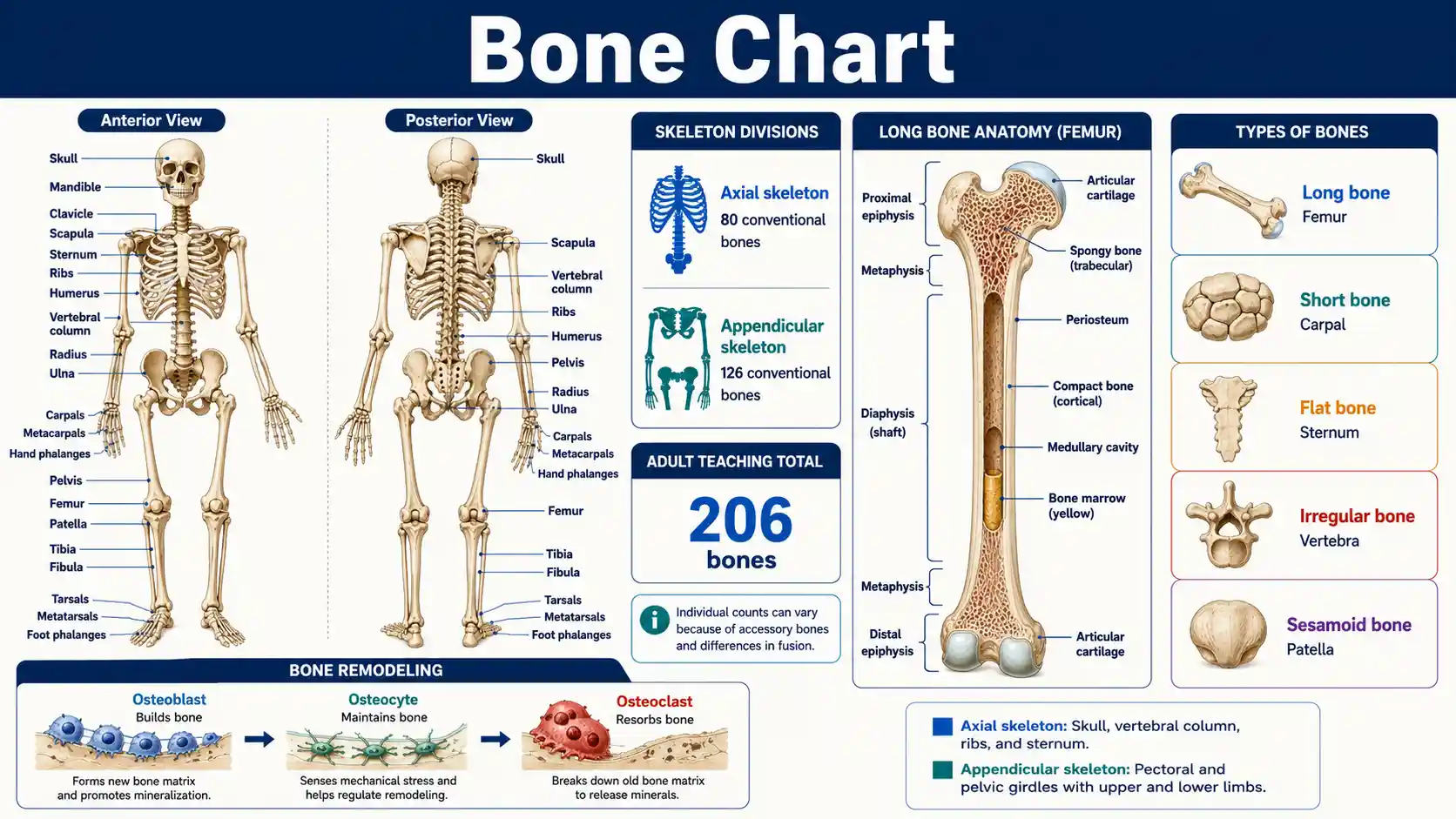
What does the human skeleton contain?
The standard adult skeleton contains 206 bones: 80 in the axial skeleton and 126 in the appendicular skeleton. Bone is living connective tissue that supports movement, protects organs, stores minerals, and houses marrow.
The OpenStax skeletal-system overview explains the conventional adult count and the anatomical boundary between the axial and appendicular divisions.
Adult teaching count
206 bones
The familiar count is a convention; accessory bones and fusion patterns can change an individual total.
Central division
80 axial bones
The skull, vertebral column, ribs, sternum, hyoid, and auditory ossicles form the axial skeleton.
Limb division
126 appendicular bones
The shoulder and pelvic girdles connect the upper and lower limbs to the central skeleton.
Living tissue
Constant remodeling
Osteoblasts build matrix, osteoclasts resorb bone, and osteocytes help maintain and sense the tissue.
Essential bone and skeleton questions
These concise answers cover the anatomy facts users most often need before reading the detailed charts.
How many bones does an adult have?
The conventional adult skeleton contains 206 bones, although normal anatomical variation can change the exact count.
What are the two main skeleton divisions?
The axial skeleton forms the central axis, while the appendicular skeleton contains the limbs and their girdles.
How many bones are axial?
The conventional adult axial skeleton contains 80 bones.
How many bones are appendicular?
The conventional adult appendicular skeleton contains 126 bones.
What is the longest bone?
The femur is the longest and strongest bone in the human body.
What is the smallest bone?
The stapes in the middle ear is generally recognized as the smallest human bone.
Are teeth bones?
No. Teeth are mineralized organs with enamel and dentin, but anatomy does not classify them as bones.
Is bone alive?
Yes. Bone contains living cells, vessels, nerves, collagen, minerals, and marrow.
Where are blood cells produced?
Many blood cells are produced in red bone marrow through hematopoiesis.
What is compact bone?
Compact bone is dense cortical tissue that provides strength and forms much of the outer bone wall.
What is spongy bone?
Spongy bone is a trabecular network that distributes stress and contains marrow spaces.
Can a chart diagnose a fracture?
No. Fracture diagnosis depends on the injury, examination, circulation and nerve checks, and imaging when needed.
Human Skeletal System Overview
The conventional adult count divides the skeleton into axial and appendicular regions. Individual totals can vary because of accessory bones, fused segments, or anatomical variation.
Swipe horizontally inside the table to view every column.
| Division | Main regions | Conventional adult count | Primary roles | Representative bones |
|---|---|---|---|---|
| Axial skeleton | Head, neck, vertebral column, and thoracic cage | 80 bones — Axial count | Supports the central axis and protects the brain, spinal cord, heart, and lungs | Skull, hyoid, vertebrae, ribs, sternum |
| Appendicular skeleton | Shoulder girdles, upper limbs, pelvic girdle, and lower limbs | 126 bones — Appendicular count | Provides attachment and leverage for limb movement and weight bearing | Clavicle, scapula, humerus, radius, ulna, hip bone, femur, tibia |
| Whole adult skeleton | Axial plus appendicular skeleton | Commonly stated as 206 bones — Conventional total | Support, movement, protection, mineral storage, and marrow functions | All named bones and paired limb elements |
The 206-bone total is a standard teaching convention, not an absolute count for every person.
- • Children have more separate skeletal elements because many bones later fuse during growth.
- • Teeth are not bones and are not included in the standard skeletal count.
- • Sesamoid and sutural bones can vary among individuals, which can change a detailed count.
Download or export
Core anatomy map
The skeleton combines regional anatomy with living tissue
Bone names describe location, while tissue terms describe internal organization. One bone can contain compact cortex, spongy trabeculae, marrow, cartilage, vessels, nerves, and remodeling cells.
Axial skeleton
80 bones
Skull, associated head bones, vertebral column, ribs, and sternum form the central body axis.
Appendicular skeleton
126 bones
Shoulder girdles, upper limbs, pelvic girdle, and lower limbs support mobility and load transfer.
Compact bone
Dense cortex
Compact bone resists bending and compression and forms much of the shaft wall in long bones.
Spongy bone
Trabecular network
Spongy bone distributes stress through trabeculae and contains marrow spaces.
The familiar total of 206 is a teaching convention. Accessory sesamoid or sutural bones and differences in fusion can change an individual count.
Axial Skeleton Bone Count by Region
The axial skeleton forms the body’s central axis. Its conventional 80-bone total includes the skull, associated head bones, vertebral column, ribs, and sternum.
Swipe horizontally inside the table to view every column.
| Region | Bones included | Count | Key protection or support role |
|---|---|---|---|
| Cranium | Frontal, parietal, temporal, occipital, sphenoid, and ethmoid bones | 8 | Encloses and protects the brain |
| Facial skeleton | Maxillae, zygomatic, nasal, lacrimal, palatine, inferior nasal conchae, vomer, and mandible | 14 | Shapes the face and supports the eyes, nose, and oral cavity |
| Auditory ossicles | Malleus, incus, and stapes in each middle ear | 6 | Transmit sound vibrations |
| Hyoid | Single U-shaped bone in the anterior neck | 1 | Supports the tongue and provides muscle attachment |
| Vertebral column | 24 vertebrae plus sacrum and coccyx as adult bones | 26 — Adult vertebral count | Supports the trunk and protects the spinal cord |
| Thoracic cage | 12 pairs of ribs plus the sternum | 25 | Protects thoracic organs and assists breathing mechanics |
| Axial total | All rows above | 80 — Axial total | Forms the central support and protection framework |
The adult sacrum and coccyx are counted as one bone each after their component vertebrae fuse.
- • The skull contains 22 cranial and facial bones under the standard convention.
- • The six auditory ossicles and the hyoid are associated with the head but counted separately from the 22 skull bones.
- • Rib number and vertebral segmentation can vary in some individuals.
Download or export
Shoulder Girdle and Upper Limb Bone Count
Each upper limb and its shoulder attachment contain 32 bones, producing 64 bones across both sides.
Swipe horizontally inside the table to view every column.
| Region | Bone or group | Per side | Both sides | Main function |
|---|---|---|---|---|
| Pectoral girdle | Clavicle | 1 | 2 | Braces the shoulder away from the trunk and transfers force |
| Pectoral girdle | Scapula | 1 | 2 | Provides a mobile platform and broad muscle attachment |
| Arm | Humerus | 1 | 2 | Forms the shoulder and elbow joints |
| Forearm | Radius | 1 | 2 | Rotates with the hand and bears more wrist load on the thumb side |
| Forearm | Ulna | 1 | 2 | Forms the main hinge with the humerus at the elbow |
| Wrist | Carpals | 8 | 16 — Both wrists | Provide flexible wrist motion |
| Hand | Metacarpals | 5 | 10 | Form the palm |
| Fingers | Phalanges | 14 | 28 | Provide digit movement and grasp |
| Upper-limb total | All girdle and free-limb bones | 32 | 64 — Upper-limb total | Supports positioning, reach, manipulation, and load transfer |
The thumb has two phalanges; each other finger has three.
- • The shoulder girdle includes the clavicle and scapula on each side.
- • The radius lies on the thumb side in anatomical position; the ulna lies on the little-finger side.
- • Carpal bones are eight short bones arranged in two rows.
Download or export
Pelvic Girdle and Lower Limb Bone Count
Each lower limb and its pelvic attachment contain 31 bones, producing 62 bones across both sides.
Swipe horizontally inside the table to view every column.
| Region | Bone or group | Per side | Both sides | Main function |
|---|---|---|---|---|
| Pelvic girdle | Hip bone | 1 | 2 | Transfers body weight to the lower limb and protects pelvic organs |
| Thigh | Femur | 1 | 2 | Supports body weight and forms the hip and knee joints |
| Knee | Patella | 1 | 2 | Protects the anterior knee and improves quadriceps leverage |
| Leg | Tibia | 1 | 2 | Main weight-bearing bone below the knee — Primary leg weight bearer |
| Leg | Fibula | 1 | 2 | Provides ankle stability and muscle attachment |
| Ankle and rearfoot | Tarsals | 7 | 14 | Transfer load and permit foot motion |
| Midfoot and forefoot | Metatarsals | 5 | 10 | Support the arches and transmit push-off forces |
| Toes | Phalanges | 14 | 28 | Assist balance and propulsion |
| Lower-limb total | All girdle and free-limb bones | 31 | 62 — Lower-limb total | Supports standing, balance, walking, running, and jumping |
The great toe has two phalanges; each other toe usually has three.
- • Each adult hip bone forms from fused ilium, ischium, and pubis components.
- • The femur is the longest and strongest bone in the human body.
- • The tibia bears substantially more body weight than the fibula.
Download or export
Why the adult bone count can vary
The conventional count treats the adult sacrum, coccyx, and sternum as single bones and excludes teeth. Accessory sutural bones, extra sesamoids, rib variants, or differences in fusion can produce a different individual total without changing the basic regional map.
Bone Shape Classification and Examples
Bones are commonly classified by overall shape. Shape relates to mechanics but does not fully describe internal structure or function.
Swipe horizontally inside the table to view every column.
| Shape class | General description | Examples | Typical mechanical role |
|---|---|---|---|
| Long bones — Shape, not absolute size | Longer than they are wide, usually with a shaft and two ends | Femur, humerus, radius, ulna, metacarpals, phalanges | Leverage, movement, and weight transfer |
| Short bones | Roughly cube-shaped with dimensions of similar scale | Carpals and tarsals | Stability with limited gliding motion |
| Flat bones | Thin, often curved plates with compact layers around spongy bone | Cranial bones, sternum, ribs, scapula | Protection and broad muscle attachment |
| Irregular bones | Complex shapes that do not fit the other main groups | Vertebrae, sphenoid, facial bones | Protection, support, and specialized attachment |
| Sesamoid bones | Develop within certain tendons near joints | Patella; variable small sesamoids in hands and feet — Variable sesamoids | Reduce tendon friction and alter mechanical leverage |
| Sutural bones | Small accessory bones within cranial sutures | Variable wormian bones | Anatomical variation rather than a required skeletal element |
Classification uses overall morphology. A “long bone” can be small, as shown by the phalanges.
- • The patella is the largest sesamoid bone.
- • Not every person has the same number of accessory sesamoid or sutural bones.
- • Some anatomy sources also describe pneumatic bones that contain air-filled spaces, especially in the skull.
Download or export
Long Bone Anatomy and Tissue Layers
A long bone such as the femur shows the major gross structures used to describe bone growth, joint surfaces, marrow spaces, and tissue organization.
Swipe horizontally inside the table to view every column.
| Structure | Location | Main role | Important distinction |
|---|---|---|---|
| Diaphysis | Tubular shaft between the ends | Provides leverage and surrounds the medullary cavity | Contains a thick compact-bone wall in a typical long bone |
| Epiphysis | Expanded proximal or distal end | Forms joint surfaces and distributes load | Contains abundant spongy bone beneath a compact shell |
| Metaphysis | Transition between shaft and epiphysis | Transfers force and contains the growth region in developing bone | Includes the epiphyseal plate before skeletal maturity |
| Articular cartilage | Covers bone where an epiphysis meets another bone | Reduces friction and absorbs compressive load | It is hyaline cartilage, not periosteum — Not periosteum |
| Periosteum | Covers most outer bone surfaces | Carries vessels and nerves and supports attachment, growth, and repair | Absent over articular cartilage |
| Endosteum | Lines internal bone surfaces and marrow spaces | Supports remodeling and repair | Thin cellular lining rather than a thick structural layer |
| Compact bone | Dense outer cortex and much of the shaft wall | Resists bending and compression | Organized into osteons in many cortical regions |
| Spongy bone | Interior of epiphyses and many flat or irregular bones | Distributes stress through trabeculae and houses marrow spaces | Also called cancellous or trabecular bone |
| Medullary cavity | Central cavity of the diaphysis | Contains marrow and reduces unnecessary mass | Often contains more yellow marrow in adults |
| Epiphyseal plate or line | Between epiphysis and metaphysis | Plate permits length growth before maturity | Cartilage plate becomes a bony line after growth ends — Growth-stage distinction |
Bone anatomy differs by bone shape, age, and skeletal region; the classic long-bone model is a teaching framework.
- • Red marrow produces blood cells; yellow marrow contains more adipose tissue.
- • Compact and spongy bone are both mineralized osseous tissue, arranged differently for mechanical demands.
- • Bone contains blood vessels and nerves and can generate pain when injured.
Download or export
Bone is mineralized, vascular, living connective tissue
The OpenStax bone-structure reference describes the periosteum, endosteum, compact cortex, spongy trabeculae, marrow cavity, growth plate, osteons, and remodeling cells used throughout the charts below.
Browser-only educational tool
Compare two major bones
Select two bones to compare their skeletal division, region, shape, pairing, articulations, function, and landmark. No information leaves the browser.
Appendicular skeleton
Femur
Thigh · Long bone
- Division
- Appendicular skeleton
- Region
- Thigh
- Shape
- Long bone
- Pairing
- Paired
- Major articulations
- Hip bone, tibia, and patella
- Primary role
- Bears weight and provides powerful leverage between hip and knee.
- Useful landmark
- Head, neck, greater trochanter, linea aspera, and condyles
Appendicular skeleton
Scapula
Pectoral girdle · Flat bone
- Division
- Appendicular skeleton
- Region
- Pectoral girdle
- Shape
- Flat bone
- Pairing
- Paired
- Major articulations
- Clavicle and humerus
- Primary role
- Provides a mobile base and extensive muscle attachment for the shoulder.
- Useful landmark
- Spine, acromion, coracoid process, and glenoid cavity
Bone Cells and Remodeling Functions
Bone is living tissue. Specialized cells build, maintain, sense, and remove mineralized matrix throughout life.
Swipe horizontally inside the table to view every column.
| Cell type | Main function | Typical location or origin | How it relates to remodeling |
|---|---|---|---|
| Osteogenic cell | Divides and differentiates into osteoblasts | Deep periosteum and marrow-associated surfaces | Supplies bone-forming precursor cells |
| Osteoblast | Produces osteoid and supports mineralization — Bone formation | Active bone-forming surfaces | Builds new bone matrix |
| Osteocyte | Maintains matrix and senses mechanical loading | Lacunae within mineralized bone | Coordinates local responses through cellular networks |
| Osteoclast | Resorbs mineralized bone — Bone resorption | Bone surfaces at resorption sites | Removes old, damaged, or unneeded tissue |
| Bone-lining cell | Covers inactive bone surfaces and regulates exchange | Quiescent periosteal and endosteal surfaces | Helps control access to the bone surface |
| Remodeling unit | Couples resorption with formation | Localized sites throughout the skeleton | Replaces microdamaged tissue and helps maintain mineral balance |
Remodeling is coordinated rather than a simple one-cell process. Hormones, loading, nutrition, age, and disease influence the balance.
- • Osteoclasts arise from the monocyte-macrophage lineage, unlike osteoblasts and osteocytes.
- • Osteoblasts can become osteocytes when surrounded by the matrix they produced.
- • Osteoporosis develops when bone loss and structural weakening outpace effective replacement over time.
Download or export
Common Bone Markings and Anatomical Terms
Bone markings describe joint surfaces, attachment points, depressions, and openings. The same term can appear on different bones.
Swipe horizontally inside the table to view every column.
| Marking | Meaning | Typical role | Example |
|---|---|---|---|
| Head | Rounded articular end carried on a narrower region | Forms a joint | Head of the femur |
| Condyle | Rounded articular prominence | Forms a joint and distributes load | Occipital condyle |
| Facet | Small, flat articular surface | Permits gliding or guides motion | Articular facet of a vertebra |
| Process | General projection from a bone | Attachment or articulation | Transverse process of a vertebra |
| Crest | Prominent ridge | Muscle or ligament attachment | Iliac crest — Palpable landmark |
| Spine | Sharp, slender projection | Attachment or landmark | Spine of the scapula |
| Tubercle | Small rounded projection | Tendon or ligament attachment | Greater tubercle of the humerus |
| Tuberosity | Large roughened projection | Strong muscle or ligament attachment | Tibial tuberosity |
| Fossa | Shallow depression | Receives a structure or creates space | Olecranon fossa |
| Foramen | Hole through a bone | Allows passage of nerves or vessels — Neurovascular passage | Foramen magnum |
| Canal or meatus | Tube-like passage | Allows passage through bone | External acoustic meatus |
| Sinus | Air-filled cavity within a bone | Reduces mass and contributes to resonance or conditioning of air | Frontal sinus |
Anatomical names should be interpreted on the specific bone and in the stated orientation.
- • A bone marking is a surface feature, not a separate bone.
- • Projection size often reflects attachment forces, but appearance also varies among individuals.
- • Foramina and canals commonly transmit nerves and blood vessels.
Download or export
Bone strength is more than bone size or mineral amount
Bone strength reflects mass, architecture, material quality, geometry, remodeling, prior injury, and load. The NIAMS bone education resource explains that growth plates support childhood length growth and that osteoblasts and osteoclasts continually renew bone tissue.
Anatomy varies
Accessory bones, fusion patterns, landmarks, and joint shapes are not identical in every person.
Images use orientation
Anterior, posterior, medial, lateral, proximal, and distal labels depend on anatomical position.
Symptoms need assessment
Pain location alone cannot distinguish fracture, joint injury, tendon injury, infection, or another cause.
Bone Injury Signs and Appropriate Next Steps
A chart cannot diagnose a fracture. Mechanism, pain, deformity, circulation, nerve function, examination, and imaging determine the next step.
Swipe horizontally inside the table to view every column.
| Finding | Possible concern | Suggested response | Why it matters |
|---|---|---|---|
| Open wound with visible bone or bone-end exposure | Open fracture | Call emergency services and cover loosely with a clean dressing; do not push tissue back — Emergency response | High risk of bleeding, contamination, and tissue injury |
| Limb is pale, cold, numb, weak, or has no detectable pulse | Blood-vessel or nerve compromise | Seek emergency care immediately — Emergency response | Delayed circulation can threaten the limb |
| Obvious deformity after major trauma | Displaced fracture or dislocation | Immobilize in the position found and seek urgent assessment | Movement can worsen soft-tissue or neurovascular injury |
| Severe neck or back pain after trauma with weakness or numbness | Spinal injury | Avoid unnecessary movement and call emergency services | The spinal cord or nerve roots may be at risk |
| Persistent focal bone pain, swelling, or inability to bear weight | Possible fracture or significant soft-tissue injury | Arrange prompt clinical assessment and imaging when advised | Some fractures are not visibly deformed |
| Minor pain that improves with rest and normal function remains possible | Lower-risk injury, but not proof of no fracture | Monitor and seek care if pain, swelling, or function worsens or persists | Stress and nondisplaced fractures can be subtle |
Emergency actions depend on local services and the person’s condition. Do not delay urgent care to compare symptoms with a web chart.
- • Do not straighten a severely deformed limb unless trained emergency guidance directs you to do so.
- • Children, older adults, people with osteoporosis, and people taking medicines that affect bone may fracture with less force.
- • A normal-looking limb can still contain a fracture.
Download or export
Frequently asked questions
How many bones are in the adult human body?
The conventional adult skeleton contains 206 bones. Individual counts can vary because accessory bones, fused segments, or anatomical variants are not identical in everyone.
Why do babies have more bones than adults?
Babies have more separate skeletal elements because many fuse during growth. Fusion reduces the conventional count as the skeleton matures.
What is the difference between the axial and appendicular skeleton?
The axial skeleton forms the central body axis, while the appendicular skeleton includes the limb bones and the girdles that attach them.
How many bones are in the axial skeleton?
The conventional adult axial skeleton contains 80 bones, including the skull, hyoid, auditory ossicles, vertebral column, ribs, and sternum.
How many bones are in the appendicular skeleton?
The conventional adult appendicular skeleton contains 126 bones in the shoulder girdles, upper limbs, pelvic girdle, and lower limbs.
What is the longest bone in the body?
The femur is the longest bone in the human body. It extends from the hip to the knee and carries substantial loads.
What is the smallest bone in the body?
The stapes in the middle ear is generally recognized as the smallest human bone. It transmits vibrations toward the inner ear.
Are teeth bones?
No. Teeth and bones are mineralized structures, but teeth have enamel and dentin and are not classified as bones.
Is bone living tissue?
Yes. Bone contains living cells, blood vessels, nerves, collagen, and mineralized matrix, and it continuously remodels.
What is the difference between compact and spongy bone?
Compact bone forms dense cortical tissue, while spongy bone forms a trabecular network with marrow spaces. Most bones contain both.
What do osteoblasts and osteoclasts do?
Osteoblasts build bone matrix, while osteoclasts resorb mineralized bone. Healthy remodeling coordinates both activities.
Where are blood cells made?
Many blood cells are produced in red bone marrow through hematopoiesis. The distribution of active red marrow changes with age.
What is a growth plate?
A growth plate is hyaline cartilage near the end of a growing long bone. It permits length growth before it is replaced by bone.
Can a bone heal after a fracture?
Bone can repair a fracture by forming new tissue, but alignment, blood supply, stability, age, and injury severity affect healing.
Does a normal X-ray rule out every fracture?
No. Some stress, nondisplaced, or very early fractures can be difficult to see, so persistent focal symptoms may need reassessment or other imaging.
What symptoms after a bone injury need emergency care?
An open wound, absent pulse, pale or cold limb, new numbness or weakness, major deformity, uncontrolled bleeding, or possible spinal injury needs emergency care.
Sources
These anatomy, physiology, histology, and skeletal-health references support the bone counts, regional classifications, tissue descriptions, remodeling functions, and interpretation limits used on this page.
OpenStax — Divisions of the Skeletal System
https://openstax.org/books/anatomy-and-physiology-2e/pages/7-1-divisions-of-the-skeletal-system
Describes the conventional 206-bone adult skeleton, including 80 axial bones and 126 appendicular bones.
OpenStax — Functions of the Skeletal System
https://openstax.org/books/anatomy-and-physiology-2e/pages/6-1-the-functions-of-the-skeletal-system
Summarizes support, movement, organ protection, mineral storage, fat storage, and blood-cell production.
OpenStax — Bone Structure
https://openstax.org/books/anatomy-and-physiology/pages/6-3-bone-structure
Explains long-bone anatomy, compact and spongy bone, bone cells, growth plates, periosteum, endosteum, and bone markings.
NCBI Bookshelf, StatPearls — Anatomy, Bones
https://www.ncbi.nlm.nih.gov/books/NBK537199/
Reviews bone anatomy, mineralized matrix, osteons, osteoblasts, osteocytes, and normal variation in adult bone count.
National Institute of Arthritis and Musculoskeletal and Skin Diseases — Learning About Bones
https://www.niams.nih.gov/health-topics/educational-resources/health-lesson-learning-about-bones
Explains growth plates, bone remodeling, osteoblasts, osteoclasts, and the fusion of many childhood bones.
MedlinePlus, U.S. National Library of Medicine — Osteoporosis
https://medlineplus.gov/osteoporosis.html
Explains that bone is living tissue and that reduced bone mass or altered structure can increase fracture risk.