Biology · Human Anatomy
Human Organ Chart for Locations, Systems, and Functions
Locate major human organs, compare their body systems and functions, identify body cavities, trace material pathways, and understand how organs work together.
An organ map is educational, not diagnostic. Pain location, fullness, tenderness, or another symptom cannot reliably identify the affected organ without clinical assessment.
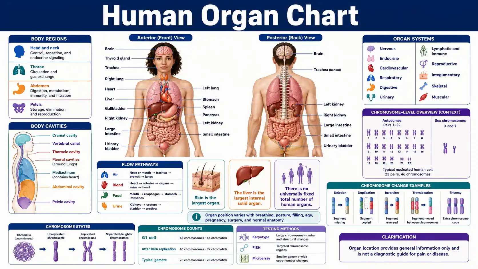
What are the major organs in the human body?
Major organs include the brain, heart, lungs, liver, stomach, intestines, pancreas, spleen, kidneys, urinary bladder, skin, and endocrine and reproductive organs. Each organ contains several tissues and works through vessels, nerves, ducts, membranes, or chemical signals.
The National Cancer Institute definition describes an organ as a body part made of cells and tissues that performs a specific function.
Definition
Several tissues, one function
An organ combines multiple tissue types into a structure that performs one or more specialized functions.
Systems framework
11 commonly taught systems
Introductory anatomy usually groups organs into eleven interacting systems, although other classifications exist.
Largest organ
Skin
Skin is generally recognized as the largest organ; the liver is the largest internal solid organ.
Counting limit
No universal total
Organ totals vary with definitions, paired structures, named segments, glands, and counting conventions.
Essential human organ questions
These concise answers establish the definitions, locations, and limitations needed to use the detailed organ charts correctly.
What is an organ?
An organ is a structure made from multiple tissue types that work together to perform specialized functions.
How many organs does the human body have?
No universally accepted total exists because anatomy sources use different definitions and counting conventions.
What is the largest organ?
Skin is generally recognized as the largest organ by surface area and mass.
What is the largest internal organ?
The liver is the largest internal solid organ in a typical adult.
Where is the heart?
The heart lies in the mediastinum between the lungs, behind the sternum, and above the diaphragm.
Where are the kidneys?
The kidneys lie high on the posterior abdominal wall behind the peritoneum, with the right usually slightly lower.
Can one organ belong to two systems?
Yes. The pancreas has digestive and endocrine roles, while the kidneys have urinary and endocrine functions.
Is blood an organ?
No. Blood is a specialized connective tissue that circulates through the cardiovascular system.
Is skin an organ?
Yes. Skin combines several tissue types and performs barrier, sensory, immune, temperature, and metabolic functions.
What does paired organ mean?
A paired organ normally occurs on both sides, although the right and left members may differ in shape or position.
Do organs remain fixed in one position?
No. Breathing, posture, filling, growth, pregnancy, surgery, and anatomical variation can shift organ relationships.
Can organ location identify the cause of pain?
No. Pain can be referred, and several organs, vessels, muscles, nerves, or membranes can cause symptoms in one region.
Major Human Organs, Locations, and Core Functions
This quick-reference chart identifies major organs by usual location, principal system, and core function. Many organs contribute to more than one system.
Swipe horizontally inside the table to view every column.
| Organ | Usual location | Principal system | Core function | Paired or unpaired |
|---|---|---|---|---|
| Brain | Cranial cavity | Nervous | Integrates sensation, movement, cognition, autonomic control, and behavior | Unpaired |
| Heart | Mediastinum in the thoracic cavity | Cardiovascular | Pumps blood through pulmonary and systemic circuits | Unpaired |
| Lungs | Right and left pleural cavities | Respiratory | Exchange oxygen and carbon dioxide between air and blood | Paired |
| Liver | Right upper abdomen below the diaphragm | Digestive and metabolic | Processes nutrients, makes bile and plasma proteins, and modifies many substances | Unpaired |
| Stomach | Left upper abdomen | Digestive | Stores, mixes, and begins chemical digestion of food | Unpaired |
| Small intestine | Central and lower abdomen | Digestive | Completes much digestion and absorbs most nutrients | Unpaired continuous tube |
| Large intestine | Frames much of the abdominal cavity | Digestive | Absorbs water and electrolytes and forms and stores feces | Unpaired continuous tube |
| Pancreas | Upper posterior abdomen behind the stomach | Digestive and endocrine — Two-system organ | Releases digestive enzymes and hormones including insulin and glucagon | Unpaired |
| Spleen | Left upper abdomen | Lymphatic and immune | Filters blood, supports immune responses, and removes aging blood cells | Unpaired |
| Kidneys | Posterior upper abdomen, retroperitoneal | Urinary | Filter blood, form urine, regulate fluid and electrolytes, and release hormones | Paired |
| Urinary bladder | Pelvic cavity | Urinary | Stores urine before urination | Unpaired |
| Skin | External body surface | Integumentary | Forms a protective barrier and supports temperature control, sensation, and vitamin D production | One continuous organ — Continuous external organ |
Locations describe standard anatomical position. Actual shape, level, and relationship vary with body size, breathing, posture, organ filling, development, and anatomy.
- • There is no single universally accepted total number of human organs because definitions and counting conventions differ.
- • An organ may belong to several functional systems; the pancreas is a common digestive and endocrine example.
- • Tubular organs such as the intestine have named segments but can be discussed as one continuous organ or multiple regions depending on context.
Download or export
Regional anatomy map
Organ location is described by regions, cavities, and relationships
A useful organ map combines body region with depth, cavity, membrane, neighboring structures, blood supply, ducts, nerves, and function. One flat diagram cannot show every three-dimensional relationship.
Head and neck
Brain and endocrine control
The brain, pituitary, thyroid, parathyroids, airway, and upper digestive tract coordinate control, sensation, breathing, and swallowing.
Thorax
Heart, lungs, and mediastinum
The lungs occupy separate pleural cavities, while the heart and several connecting structures lie in the central mediastinum.
Abdomen
Digestion, metabolism, and filtration
The liver, stomach, pancreas, spleen, intestines, and kidneys exchange materials through ducts, vessels, nerves, and the peritoneal or retroperitoneal spaces.
Pelvis
Storage, elimination, and reproduction
The urinary bladder, rectum, internal reproductive organs, vessels, nerves, and pelvic floor share a compact region and influence one another mechanically.
Cavity
The space or compartment that contains or surrounds an organ, such as the cranial, pleural, pericardial, abdominal, or pelvic region.
Relationship
Directional terms explain whether one organ is superior, inferior, medial, lateral, anterior, posterior, superficial, or deep to another.
Connection
Organs coordinate through blood vessels, nerves, ducts, airways, lumens, lymphatics, ligaments, membranes, and chemical signals.
Eleven Human Organ Systems and Their Main Organs
An introductory anatomy framework commonly groups the body into eleven interacting organ systems. These boundaries are useful for study but do not isolate organs from one another.
Swipe horizontally inside the table to view every column.
| Organ system | Representative organs and structures | Primary functions | Important overlap |
|---|---|---|---|
| Integumentary | Skin, hair, nails, sweat glands, sebaceous glands | Protection, sensation, temperature regulation, and vitamin D production | Works with immune, nervous, and circulatory systems |
| Skeletal | Bones, cartilage, ligaments, bone marrow | Support, protection, leverage, mineral storage, and blood-cell production | Works closely with muscular and hematopoietic systems |
| Muscular | Skeletal muscles and associated tendons | Movement, posture, joint stabilization, and heat production | Cardiac and smooth muscle are located in other organ systems |
| Nervous | Brain, spinal cord, peripheral nerves, sensory organs | Rapid communication, integration, sensation, and motor control | Closely coordinates endocrine and muscular activity |
| Endocrine | Pituitary, thyroid, parathyroids, adrenals, pancreatic islets, ovaries or testes | Hormonal regulation of growth, metabolism, stress, fluid balance, and reproduction | Many endocrine organs also belong to another system |
| Cardiovascular | Heart, arteries, veins, capillaries, blood | Transports gases, nutrients, wastes, hormones, heat, and immune cells | Connects functionally with every organ system — Whole-body connection |
| Lymphatic and immune | Lymphatic vessels, lymph nodes, spleen, thymus, tonsils | Returns tissue fluid and supports immune surveillance and responses | Closely linked with blood and mucosal organs |
| Respiratory | Nose, pharynx, larynx, trachea, bronchi, lungs | Ventilation, gas exchange, acid-base support, voice, and airway defense | Works directly with cardiovascular circulation |
| Digestive | Mouth, esophagus, stomach, intestines, liver, gallbladder, pancreas | Digestion, nutrient absorption, water recovery, and elimination of solid waste | Liver and pancreas also perform major metabolic or endocrine roles |
| Urinary | Kidneys, ureters, urinary bladder, urethra | Urine formation, waste removal, fluid and electrolyte control, and acid-base regulation | Kidneys also have endocrine functions |
| Reproductive | Ovaries, uterine tubes, uterus, vagina; or testes, ducts, prostate, penis — Sex-specific anatomy | Produces gametes and sex hormones and supports reproduction | Anatomy varies, and several organs also have endocrine roles |
System names follow a common introductory anatomy convention. Some curricula combine or subdivide systems differently.
- • Organ systems maintain homeostasis through continuous interaction rather than independent operation.
- • Blood is a connective tissue, while the heart and vessels are organs of the cardiovascular system.
- • The reproductive system contains sex-specific structures; not every listed organ is present in every body.
Download or export
Organ systems are study frameworks, not isolated machines
The OpenStax structural-organization reference presents eleven commonly taught systems. Blood carries respiratory gases and hormones, nerves coordinate organ activity, and kidneys, lungs, skin, and digestive organs jointly regulate the internal environment.
Body Cavities and the Organs They Contain
Body cavities organize internal anatomy and allow organs to move, expand, and remain protected. Serous membranes reduce friction around several thoracic and abdominopelvic organs.
Swipe horizontally inside the table to view every column.
| Cavity or space | Major boundaries | Representative contents | Key membrane or relationship |
|---|---|---|---|
| Cranial cavity | Inside the skull | Brain, meninges, vessels, and cranial nerve roots | Part of the dorsal body cavity |
| Vertebral canal | Within the vertebral column | Spinal cord, meninges, nerve roots, vessels, and cerebrospinal fluid spaces | Continuous with the cranial cavity |
| Thoracic cavity | Chest wall above the diaphragm | Pleural cavities and mediastinum | Separated from the abdominopelvic cavity by the diaphragm |
| Right pleural cavity | Around the right lung | Right lung and pleural fluid space | Visceral and parietal pleura form a low-friction interface |
| Left pleural cavity | Around the left lung | Left lung and pleural fluid space | Separate from the right pleural cavity |
| Mediastinum | Central thoracic compartment between lungs | Heart, great vessels, trachea, esophagus, thymus, nerves, and lymphatics — Central thoracic organs | Contains the pericardial cavity around the heart |
| Abdominal cavity | Below the diaphragm and above the pelvic brim | Liver, stomach, spleen, pancreas, intestines, and other structures | Many organs are intraperitoneal; others are retroperitoneal |
| Pelvic cavity | Within the bony pelvis | Urinary bladder, rectum, internal reproductive organs, vessels, and nerves | Continuous superiorly with the abdominal cavity |
| Peritoneal cavity | Potential space between parietal and visceral peritoneum | Thin lubricating fluid, not the solid abdominal organs themselves — Potential-space distinction | Organs may be intraperitoneal or retroperitoneal |
A potential space normally contains only a thin fluid film. It should not be confused with the organs enclosed or covered by its membranes.
- • The dorsal cavity includes the cranial cavity and vertebral canal.
- • The ventral cavity includes thoracic and abdominopelvic subdivisions.
- • The kidneys and most of the pancreas lie primarily behind the peritoneum in the retroperitoneal space.
Download or export
Paired, Unpaired, and Midline Organ Examples
Pairing describes whether a structure normally occurs on both sides. It does not mean the two organs are perfectly identical or equally positioned.
Swipe horizontally inside the table to view every column.
| Pattern | Examples | Location clue | Important variation |
|---|---|---|---|
| Paired organs | Lungs, kidneys, adrenal glands, ovaries or testes, eyes, ears | One on the right and one on the left | Paired organs may differ in size, shape, level, or lobes — Paired does not mean identical |
| Unpaired organs | Heart, liver, stomach, spleen, pancreas, urinary bladder | Single organ with a characteristic position | An unpaired organ can cross the midline or have right and left parts |
| Midline organs | Brain, spinal cord, trachea, esophagus, uterus, prostate | Centered on or near the median plane | Some are only approximately midline or shift with filling and movement |
| Repeated segmental organs or units | Vertebrae, ribs, lymph nodes, nephrons, lung lobules | Multiple repeated structures | Not every repeated structure is considered a separate organ |
| Continuous tubular organs | Small intestine, large intestine, blood vessels, lymphatic vessels | Extend through several regions | Naming and counting depend on whether segments are treated separately — Counting convention |
“Paired” describes the common anatomical plan. Congenital absence, surgical removal, transplantation, fusion, duplication, or positional variants can change an individual pattern.
- • The right lung usually has three lobes, while the left usually has two to accommodate the heart.
- • The right kidney usually sits slightly lower than the left because of the liver.
- • The heart is unpaired but lies obliquely and mostly left of the midline rather than entirely on the left side.
Download or export
Digestive Tract Pathway and Accessory Organs
Food passes through the gastrointestinal tract. Accessory organs support digestion by producing, storing, or releasing secretions without food normally passing through them.
Swipe horizontally inside the table to view every column.
| Structure | Food passes through? | Main role | Connection to the next stage |
|---|---|---|---|
| Mouth and oral cavity | Yes | Chews food, mixes it with saliva, and begins swallowing and carbohydrate digestion | Passes the bolus to the pharynx |
| Pharynx | Yes | Directs swallowed material from the mouth toward the esophagus | Coordinates passage while the airway is protected |
| Esophagus | Yes | Moves the bolus to the stomach by peristalsis | Crosses the diaphragm and enters the stomach |
| Stomach | Yes | Stores and mixes food and begins substantial protein digestion | Releases chyme into the duodenum |
| Small intestine | Yes | Receives bile and pancreatic secretions, completes digestion, and absorbs most nutrients — Main nutrient-absorption site | Passes remaining contents to the large intestine |
| Large intestine | Yes | Absorbs water and electrolytes and compacts fecal material | Moves feces toward the rectum and anal canal |
| Liver | No — Accessory organ | Produces bile and performs nutrient processing, detoxification, synthesis, and storage | Bile reaches the small intestine through the biliary tree |
| Gallbladder | No | Stores and concentrates bile | Contracts to deliver bile into the small intestine |
| Pancreas | No | Releases digestive enzymes and bicarbonate into the duodenum | Pancreatic ducts join or approach the bile duct near the duodenum |
| Salivary glands | No | Produce saliva for lubrication, buffering, and initial digestion | Ducts empty into the mouth |
The gastrointestinal tract is a continuous lumen from mouth to anus. Accessory organs connect through ducts, vessels, nerves, or biochemical exchange.
- • The liver and pancreas have major functions beyond digestion.
- • The appendix is attached to the cecum and contains lymphoid tissue; it is not simply a useless structure.
- • The microbiome participates in digestive and metabolic functions but is not itself a human organ.
Download or export
Browser-only educational tool
Compare two human organs
Select two organs to compare their system, cavity, pairing pattern, major input and output, function, connections, and location clues. No information leaves the browser.
Cardiovascular
Heart
Pericardial cavity within the mediastinum
- Organ system
- Cardiovascular
- Cavity or region
- Pericardial cavity within the mediastinum
- Pattern
- Unpaired
- Primary function
- Generates pressure to move blood through pulmonary and systemic circulation.
- Main input
- Venous blood returning from the body and lungs
- Main output
- Blood pumped into pulmonary and systemic arteries
- Key connections
- Great veins, pulmonary trunk, pulmonary veins, aorta, coronary vessels, and conduction system
- Location clue
- Between the lungs, behind the sternum, above the diaphragm
Digestive and metabolic
Liver
Upper abdominal cavity, mostly intraperitoneal
- Organ system
- Digestive and metabolic
- Cavity or region
- Upper abdominal cavity, mostly intraperitoneal
- Pattern
- Unpaired
- Primary function
- Processes nutrients, produces bile and plasma proteins, stores substances, and modifies toxins and medicines.
- Main input
- Portal blood from digestive organs and arterial blood from the hepatic artery
- Main output
- Hepatic venous blood, bile, plasma proteins, and processed metabolites
- Key connections
- Portal vein, hepatic artery, hepatic veins, bile ducts, gallbladder, and duodenum
- Location clue
- Mostly in the right upper abdomen beneath the diaphragm
How Air, Blood, Food, and Urine Move Between Organs
These simplified pathways show the direction of major body flows. Real physiology includes branches, feedback loops, valves, pressure gradients, and microscopic exchange surfaces.
Swipe horizontally inside the table to view every column.
| Flow | Simplified organ pathway | Main exchange or transformation | Final destination or cycle |
|---|---|---|---|
| Air | Nose or mouth → pharynx → larynx → trachea → bronchi → lungs | Gas exchange occurs across alveolar-capillary membranes | Exhaled air returns through the conducting airways |
| Pulmonary blood | Right heart → pulmonary arteries → lung capillaries → pulmonary veins → left heart — Pulmonary circuit | Blood releases carbon dioxide and takes up oxygen | Oxygenated blood enters systemic circulation |
| Systemic blood | Left heart → arteries → tissue capillaries → veins → right heart | Cells exchange gases, nutrients, wastes, heat, and signals | Deoxygenated blood returns to the pulmonary circuit |
| Food and digestive contents | Mouth → esophagus → stomach → small intestine → large intestine → rectum → anus | Digestion and absorption move nutrients into blood or lymph | Unabsorbed residue leaves as feces |
| Bile | Liver → bile ducts; gallbladder storage when appropriate → common bile duct → duodenum | Bile salts help emulsify and absorb fats | Much bile salt is recycled through the liver |
| Urine | Kidneys → ureters → urinary bladder → urethra — Urinary flow | Kidneys filter and modify tubular fluid to form urine | Urine leaves the body during urination |
| Lymph | Tissue spaces → lymphatic capillaries → vessels and nodes → major ducts → veins | Returns fluid and transports immune cells and absorbed fats | Rejoins the bloodstream near the neck |
Arrows indicate the dominant direction under normal physiology. They do not show every branch or regulatory step.
- • Arteries carry blood away from the heart and veins return blood to the heart; oxygen level is not the defining distinction.
- • Pulmonary arteries carry oxygen-poor blood, while pulmonary veins carry oxygen-rich blood.
- • Kidneys receive blood through renal arteries, but urine travels through ureters, not blood vessels.
Download or export
Major Endocrine Organs and Hormone Roles
Endocrine organs release hormones into the bloodstream. Several also belong to another organ system and perform non-endocrine functions.
Swipe horizontally inside the table to view every column.
| Organ or gland | Usual location | Representative hormones | Major regulated functions |
|---|---|---|---|
| Hypothalamus | Base of the brain | Releasing and inhibiting hormones; produces oxytocin and vasopressin | Links nervous and endocrine control and regulates pituitary activity |
| Pituitary gland | Sella turcica below the hypothalamus | ACTH, TSH, GH, prolactin, LH, FSH; releases oxytocin and vasopressin | Coordinates many endocrine glands, growth, reproduction, and water balance |
| Pineal gland | Brain near the roof of the third ventricle | Melatonin | Supports circadian timing |
| Thyroid gland | Anterior neck around the trachea | Thyroid hormones and calcitonin | Metabolic rate, growth, development, and calcium-related signaling |
| Parathyroid glands | Usually on the posterior thyroid | Parathyroid hormone | Raises blood calcium through coordinated bone, kidney, and intestinal effects |
| Adrenal glands | On the superior poles of the kidneys | Cortisol, aldosterone, adrenal androgens, epinephrine, norepinephrine | Stress responses, blood pressure, electrolyte balance, and metabolism |
| Pancreatic islets — Dual-system organ | Within the pancreas | Insulin, glucagon, somatostatin, pancreatic polypeptide | Blood-glucose and nutrient regulation |
| Ovaries | Pelvic cavity | Estrogens, progesterone, inhibins | Oocyte development, reproductive cycles, pregnancy support, and sex characteristics |
| Testes | Scrotum | Testosterone and inhibin | Sperm production, reproductive function, and sex characteristics |
| Kidneys — Urinary and endocrine roles | Posterior abdomen | Renin, erythropoietin, active vitamin D-related hormone | Blood pressure, red-cell production, and calcium-phosphate balance |
Hormone lists are representative rather than exhaustive. Hormones act through receptors in target cells and are regulated by feedback loops.
- • The pancreas is both an endocrine organ and an exocrine digestive organ.
- • The kidneys are urinary organs with important endocrine functions.
- • The placenta is a temporary endocrine organ during pregnancy.
Download or export
Organ location is best learned in three dimensions
The MedlinePlus anatomy collection links organ and system illustrations from head to toe. Cross-sectional and three-dimensional views help clarify depth, overlap, paired structures, and the way organs move with breathing or filling.
Anatomical Terms Used to Locate Human Organs
Standard directional terms describe relationships in anatomical position. They are more precise than everyday labels such as “above,” “behind,” or “on the left.”
Swipe horizontally inside the table to view every column.
| Term | Meaning | Organ-location example | Common mistake to avoid |
|---|---|---|---|
| Superior | Toward the head or above another structure | The heart is superior to the diaphragm | Do not substitute “higher” when body position changes |
| Inferior | Toward the feet or below another structure | The urinary bladder is inferior to the kidneys | Inferior does not mean less important |
| Anterior | Toward the front of the body | The sternum is anterior to the heart | Anterior is defined from anatomical position |
| Posterior | Toward the back of the body | The kidneys are posterior to most abdominal digestive organs | Posterior does not always mean directly behind |
| Medial | Closer to the midline | The heart is medial to the lungs | Medial is relational, not synonymous with middle — Relational term |
| Lateral | Farther from the midline | The lungs are lateral to the heart | Right and left refer to the person, not the viewer — Patient-side convention |
| Superficial | Closer to the body surface | The skin is superficial to skeletal muscle and internal organs | A superficial organ can still have deep layers |
| Deep | Farther from the body surface | The heart is deep to the sternum | Deep is relational and does not specify a cavity |
| Proximal | Nearer the trunk or point of origin | The proximal ureter is nearer the kidney | Used most often for limbs and tubular structures |
| Distal | Farther from the trunk or point of origin | The distal esophagus is nearer the stomach | Distal does not necessarily mean lower |
| Ipsilateral | On the same side | The right kidney and right adrenal gland are ipsilateral | Does not imply direct contact |
| Contralateral | On opposite sides | The right lung is contralateral to the left kidney | Does not describe functional connection |
All directional descriptions assume standard anatomical position unless a different orientation is specified.
- • In medical images, orientation labels must be checked before interpreting right and left.
- • Cross-sectional views use axial, coronal, and sagittal planes to show organ relationships.
- • Organ position can change with breathing, posture, pregnancy, filling, development, surgery, or anatomical variation.
Download or export
Human organ anatomy has normal variation and important limits
Diagrams show a standard arrangement, but real anatomy varies. Organ size and position can change with age, breathing, posture, body composition, pregnancy, filling, congenital differences, surgery, transplantation, or disease.
No fixed organ total
Counts depend on whether paired organs, glands, named segments, and continuous structures are grouped or separated.
Images flatten depth
Front-view diagrams can hide posterior organs, membranes, vessels, ducts, and structures that overlap in three dimensions.
Symptoms overlap
Pain and other symptoms can arise from nearby tissues or be referred from a different organ or body region.
Symptoms That Need Urgent Medical Assessment
An organ chart cannot identify the cause of symptoms. Some patterns require urgent assessment because they may signal time-sensitive problems involving the brain, heart, lungs, abdomen, kidneys, or other organs.
Swipe horizontally inside the table to view every column.
| Symptom pattern | Why it can be urgent | Recommended response | Important limitation |
|---|---|---|---|
| New facial droop, arm weakness, speech difficulty, severe confusion, or sudden loss of coordination | May indicate an acute brain or blood-vessel emergency | Call emergency services immediately — Emergency action | Do not wait for symptoms to resolve or use a chart to localize the lesion |
| Chest pressure with shortness of breath, sweating, nausea, faintness, or pain spreading to the arm, jaw, or back | May indicate a heart or major-vessel emergency | Call emergency services immediately — Emergency action | Symptoms vary, and not every serious cardiac event feels the same |
| Severe breathing difficulty, blue or gray lips, inability to speak normally, or rapidly worsening wheeze | May indicate respiratory failure or severe airway obstruction | Call emergency services immediately — Emergency action | A normal-looking organ diagram cannot assess oxygenation |
| Sudden severe abdominal pain, rigid abdomen, persistent vomiting, vomiting blood, or black or bloody stool | May indicate bleeding, obstruction, perforation, inflammation, or another acute abdominal problem | Seek emergency assessment | Pain location alone cannot identify the organ involved |
| No urine with severe pain, marked swelling, confusion, or breathing difficulty | May indicate obstruction, acute kidney dysfunction, or dangerous fluid imbalance | Seek urgent medical assessment | Urine volume varies, but major sudden change with illness needs evaluation |
| Severe allergic swelling of the lips or tongue, difficulty breathing, or faintness | May indicate anaphylaxis affecting the airway and circulation | Use prescribed emergency medication if available and call emergency services | Do not delay emergency care to identify the responsible organ |
| Major trauma with deformity, uncontrolled bleeding, loss of consciousness, or severe neck, chest, abdominal, or pelvic pain | Internal organs can be injured without visible external damage | Call emergency services and avoid unnecessary movement | Only trained assessment and imaging can evaluate internal injury |
Emergency numbers and care pathways differ by country. Use the local emergency service for immediately life-threatening symptoms.
- • This list is not exhaustive and cannot replace clinical triage.
- • Children, pregnant people, older adults, and people with chronic illness may present differently.
- • When symptoms are severe, sudden, rapidly worsening, or associated with collapse, seek urgent help rather than self-diagnosing by organ location.
Download or export
Frequently asked questions
What is an organ?
An organ is a body structure made from multiple tissue types that work together to perform one or more specialized functions.
How many organs are in the human body?
There is no single universally accepted total. The count changes with definitions, whether paired structures are counted separately, and whether organ segments or glands are grouped together.
What is the largest human organ?
The skin is generally recognized as the largest organ by surface area and mass. The liver is the largest internal solid organ.
What is the largest internal organ?
The liver is the largest internal solid organ in a typical adult.
Is the brain an organ?
Yes. The brain is an organ of the nervous system composed of nervous, vascular, connective, and supporting tissues.
Is blood an organ?
No. Blood is classified as a specialized connective tissue, although it circulates through the cardiovascular organ system.
Is skin an organ?
Yes. Skin contains several tissue types and performs barrier, sensory, immune, temperature-regulating, and metabolic functions.
Can one organ belong to more than one system?
Yes. The pancreas belongs to both digestive and endocrine frameworks, and the kidneys have urinary and endocrine functions.
Which organs are paired?
Common paired organs include the lungs, kidneys, adrenal glands, eyes, ears, and ovaries or testes.
Where are the kidneys located?
The kidneys lie high on the posterior abdominal wall behind the peritoneum, with the right kidney usually slightly lower than the left.
Where is the heart located?
The heart lies in the mediastinum between the lungs, behind the sternum, above the diaphragm, and mostly left of the midline.
Where is the liver located?
Most of the liver lies in the right upper abdomen beneath the diaphragm, although it extends across the midline.
Are all internal organs called viscera?
Viscera usually refers to soft internal organs, especially those in the thoracic, abdominal, and pelvic cavities. Usage varies by context.
What is an organ system?
An organ system is a group of organs and related structures that coordinate to perform major body functions.
Do organs stay in exactly the same position?
No. Organ position changes with breathing, posture, filling, body size, age, pregnancy, surgery, and normal anatomical variation.
Can an organ chart diagnose pain?
No. Pain may be referred from another structure, and several organs, muscles, nerves, vessels, or membranes can produce symptoms in the same region.
Sources
These anatomy, physiology, terminology, body-cavity, digestive, endocrine, and urinary references support the organ locations, system relationships, pathways, and limitations used throughout the page.
OpenStax — Structural Organization of the Human Body
https://openstax.org/books/anatomy-and-physiology-2e/pages/1-2-structural-organization-of-the-human-body
Defines organs and organ systems and summarizes the eleven commonly taught human organ systems with representative structures and functions.
OpenStax — Anatomical Terminology
https://openstax.org/books/anatomy-and-physiology-2e/pages/1-6-anatomical-terminology
Explains anatomical position, directional terms, body planes, regions, and the major body cavities used to describe organ locations.
MedlinePlus — Anatomy
https://medlineplus.gov/anatomy.html
Provides NIH-supported anatomy resources and organ-system references organized from head to toe.
National Cancer Institute — Definition of Organ
https://www.cancer.gov/publications/dictionaries/cancer-terms/def/organ
Defines an organ as a body part made of cells and tissues that performs a specific function and lists familiar examples.
SEER Training Modules — Introduction to the Human Body
https://training.seer.cancer.gov/anatomy/body/
Explains the hierarchy from cells to tissues, organs, and systems and describes how several tissue types combine within an organ.
SEER Training Modules — Anatomical Terminology and Body Cavities
https://training.seer.cancer.gov/anatomy/body/terminology.html
Describes dorsal and ventral cavities and the thoracic and abdominopelvic subdivisions that contain major internal organs.
National Cancer Institute — Definition of Digestive System
https://www.cancer.gov/publications/dictionaries/cancer-terms/def/digestive-system
Lists the gastrointestinal tract and accessory digestive organs and summarizes digestion, absorption, and waste elimination.
National Cancer Institute — Definition of Endocrine System
https://www.cancer.gov/publications/dictionaries/cancer-terms/def/endocrine-system
Identifies major hormone-producing glands and organs and describes endocrine control of growth, metabolism, and reproduction.
Centers for Disease Control and Prevention — Signs and Symptoms of Stroke
https://www.cdc.gov/stroke/signs-symptoms/index.html
Lists sudden stroke warning signs and advises immediate emergency-service activation.
MedlinePlus — Heart Attack First Aid
https://medlineplus.gov/ency/article/000063.htm
Provides emergency first-aid guidance for suspected heart attack, including calling the local emergency number.
MedlinePlus — Breathing Difficulties — First Aid
https://medlineplus.gov/ency/article/000007.htm
Describes severe breathing-difficulty warning signs and the need for immediate emergency help.
MedlinePlus — Anaphylaxis
https://medlineplus.gov/anaphylaxis.html
Summarizes airway, breathing, circulation, skin, and gastrointestinal signs of a life-threatening allergic reaction.
MedlinePlus — Abdominal Pain
https://medlineplus.gov/ency/article/003120.htm
Identifies urgent abdominal symptoms such as sudden severe pain, a rigid abdomen, vomiting blood, and bloody or black stool.
National Cancer Institute — Definition of Kidney
https://www.cancer.gov/publications/dictionaries/cancer-terms/def/kidney
Summarizes kidney filtration, fluid and electrolyte regulation, blood-pressure effects, and hormone-related functions.