Biology · Hematology and Cell Biology
Blood Cell Chart for Types, Functions, Morphology, and CBC Measures
Compare red blood cells, the five major white blood cell types, and platelets. Learn how they form, what they do, how they look on a smear, and how a complete blood count describes them.
This chart is educational and cannot interpret an individual CBC. Use the reference interval printed by the testing laboratory and discuss unexpected results with a qualified clinician. Symptoms, trends, specimen quality, age, pregnancy, altitude, medicines, and other tests can change the meaning.
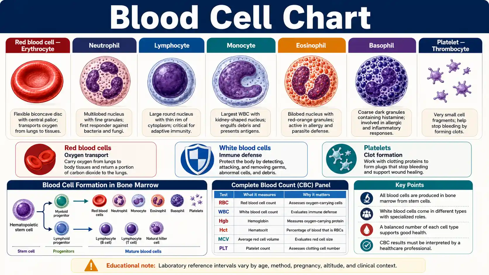
What are blood cells and what do they do?
Red blood cells transport respiratory gases, white blood cells provide immune defense, and platelets help stop bleeding. Bone marrow continually replaces these formed elements through hematopoiesis.
The MedlinePlus complete blood count guide explains that a CBC measures the number and size of blood cells and reports related measurements such as hemoglobin, hematocrit, and cell indices.
Oxygen transport
Red blood cells
Erythrocytes carry oxygen through hemoglobin and circulate as flexible biconcave discs.
Immune defense
White blood cells
Leukocytes include neutrophils, lymphocytes, monocytes, eosinophils, and basophils.
Vessel repair
Platelets
Platelets adhere and aggregate at damaged vessels and support formation of a stable clot.
Production site
Bone marrow
Hematopoietic stem and progenitor cells continuously generate new blood-cell lineages.
Essential blood cell questions
These direct answers connect cell structure and function with the most common CBC terms.
What are the main blood cell types?
The main formed elements are red blood cells, white blood cells, and platelets.
What carries oxygen in blood?
Red blood cells carry oxygen through the iron-containing protein hemoglobin.
What fights infection in blood?
White blood cells provide immune defense, but each leukocyte type performs a different role.
What helps stop bleeding?
Platelets form an initial plug and work with coagulation proteins to stabilize a clot.
Are platelets true cells?
Platelets are anucleate fragments released from large bone-marrow megakaryocytes.
Which white cell is most common?
Neutrophils are usually the most common leukocyte in adult peripheral blood.
What does a CBC measure?
A CBC measures blood-cell counts and reports hemoglobin, hematocrit, and selected cell indices.
What is a WBC differential?
A differential separates total white cells into neutrophils, lymphocytes, monocytes, eosinophils, and basophils.
What does MCV show?
MCV shows the average volume of a red blood cell and is usually reported in femtoliters.
What does hematocrit show?
Hematocrit shows the percentage of blood volume occupied by red blood cells.
Can one abnormal count diagnose a disease?
No. One blood-cell result cannot identify a cause without context, confirmation, and other findings.
Why can reference ranges differ?
Laboratories use different analyzers, methods, units, and reference populations.
Main Blood Cell Types and Their Core Functions
Whole blood contains plasma plus formed elements. The major formed elements are red blood cells, white blood cells, and platelets.
Swipe horizontally inside the table to view every column.
| Component | Alternative name | Main biological role | Key structural feature |
|---|---|---|---|
| Red blood cell | Erythrocyte or RBC | Transports oxygen through hemoglobin and contributes to carbon-dioxide transport — Oxygen transport | Mature human cells are flexible biconcave discs without nuclei |
| White blood cell | Leukocyte or WBC | Provides innate and adaptive immune defense | Nucleated cells with several distinct lineages and morphologies |
| Platelet | Thrombocyte | Forms a platelet plug and supports coagulation at damaged vessels | Small anucleate fragments released by megakaryocytes — Cell fragment |
| Plasma | Liquid blood component | Carries cells, proteins, electrolytes, nutrients, hormones, and waste | Mostly water with dissolved proteins and solutes; plasma is not a blood cell |
A formed element is a cellular or cell-derived component suspended in plasma. Platelets are fragments rather than complete nucleated cells.
- • Red blood cells are the most numerous formed element in circulating blood.
- • White blood cells include neutrophils, lymphocytes, monocytes, eosinophils, and basophils.
- • Blood function depends on both cells and plasma; a cell chart does not represent the entire composition of blood.
Download or export
How blood cells are produced and renewed
Hematopoiesis continuously replaces circulating cells. One stem-cell system produces oxygen-carrying erythrocytes, immune leukocytes, and platelet-producing megakaryocytes.
1
Stem cells divide in bone marrow
Hematopoietic stem cells generate myeloid and lymphoid progenitor lineages.
2
Precursors mature into specialized cells
Maturation changes cell size, nucleus shape, granules, proteins, and functional capacity.
3
Cells enter blood or tissues
Red cells circulate, platelets patrol vessels, and many leukocytes migrate into tissues.
4
Old or activated cells are cleared
The spleen, liver, macrophages, programmed cell death, and tissue turnover remove cells.
Blood-cell production is dynamic. Infection, inflammation, bleeding, oxygen demand, medicines, nutrition, and bone-marrow disorders can change production and release patterns.
White blood cells are specialized immune populations
The NCBI overview of blood and its cells describes red-cell oxygen transport, white-cell immune defense, and platelet clotting. A routine differential distinguishes five circulating leukocyte groups, but each group contains additional functional diversity.
Five White Blood Cell Types and Immune Roles
A white blood cell differential separates leukocytes into five major circulating types. Each lineage contributes differently to immune defense and inflammation.
Swipe horizontally inside the table to view every column.
| White cell | Group | Main role | Typical smear clue |
|---|---|---|---|
| Neutrophil — Common adult leukocyte | Granulocyte | Rapid phagocytic response to many microbes and tissue injury | Multilobed nucleus with fine pale granules |
| Lymphocyte | Agranulocyte | B-cell antibody responses, T-cell regulation and killing, and natural-killer-cell cytotoxicity | Dense round nucleus with a thin rim of cytoplasm in small cells |
| Monocyte | Agranulocyte | Phagocytosis, antigen presentation, tissue surveillance, and macrophage or dendritic-cell differentiation | Large cell with folded or kidney-shaped nucleus |
| Eosinophil | Granulocyte | Parasite defense, allergic responses, and inflammatory regulation — Allergy and parasite context | Bilobed nucleus with large orange-red granules |
| Basophil | Granulocyte | Immediate hypersensitivity and release of inflammatory mediators | Coarse dark blue-purple granules may obscure the nucleus |
Granulocytes have conspicuous cytoplasmic granules after routine staining. Agranulocytes lack the same prominent specific granules but still contain lysosomal structures.
- • Neutrophils are usually the most common white blood cell in adult peripheral blood.
- • Lymphocytes include functionally different B cells, T cells, and natural killer cells that can look similar on a routine smear.
- • Cell appearance alone does not determine function, activation state, clonality, or diagnosis.
Download or export
Blood Cell Morphology, Size, Nucleus, and Lifespan
Approximate dimensions and lifespan descriptions help compare cell biology. Values vary with preparation, maturation, activation, and source.
Swipe horizontally inside the table to view every column.
| Cell type | Approximate diameter | Nucleus | Lifespan or maturation context | Recognition clue |
|---|---|---|---|---|
| Mature red blood cell | About 7–8 µm — RBC reference size | Absent | About 120 days in typical circulation | Biconcave disc with central pallor |
| Reticulocyte | Slightly larger than a mature RBC | Absent; residual RNA remains | Completes maturation after marrow release, often within one to two days | Reticular RNA appears with supravital staining |
| Neutrophil | About 10–15 µm | Usually 2–5 lobes | Short circulation phase; survival changes after tissue entry | Fine granules and segmented nucleus |
| Small lymphocyte | About 7–10 µm | Large, round, dense | Ranges from short-lived effectors to long-lived memory cells | Thin rim of blue cytoplasm |
| Monocyte | About 15–20 µm | Indented or folded | Circulates for days before many cells enter tissues | Gray-blue cytoplasm and kidney-shaped nucleus |
| Eosinophil | About 12–17 µm | Usually bilobed | Brief blood phase with variable tissue survival | Large orange-red granules |
| Basophil | About 10–14 µm | Lobed, often obscured | Short-lived circulating granulocyte | Coarse blue-purple granules |
| Platelet | About 2–3 µm — Smallest formed element | Absent | About 7–10 days in circulation | Tiny purple-staining fragment |
µm means micrometer, equal to one-millionth of a meter. Dimensions are approximate and are not diagnostic cutoffs.
- • A standard blood smear spreads cells into a thin layer, fixes them, and applies stains that accent nuclei, cytoplasm, and granules.
- • Automated analyzers measure many features differently from manual microscopy.
- • Immature, activated, dysplastic, infected, or damaged cells may not match textbook morphology.
Download or export
Blood Cell Formation and Hematopoietic Lineages
Hematopoietic stem cells in bone marrow generate progenitors that mature into circulating blood cells and tissue immune populations.
Swipe horizontally inside the table to view every column.
| Lineage or stage | Major descendants | Main maturation site | Key signal or biological context |
|---|---|---|---|
| Hematopoietic stem cell | All blood-cell lineages | Bone marrow | Self-renewal and controlled differentiation maintain blood-cell supply |
| Common myeloid progenitor | Erythroid, megakaryocytic, granulocytic, and monocytic lineages | Bone marrow | Produces most formed elements except the main lymphoid lineages |
| Erythroid lineage | Reticulocytes and mature erythrocytes — Red-cell lineage | Bone marrow, then blood | Erythropoietin supports production when oxygen delivery is reduced |
| Megakaryocytic lineage | Megakaryocytes and platelets — Platelet source | Bone marrow | Megakaryocyte cytoplasm fragments into circulating platelets |
| Granulocytic lineage | Neutrophils, eosinophils, and basophils | Bone marrow | Granules and nuclear shape change during maturation |
| Monocytic lineage | Monocytes and many tissue macrophage or dendritic-cell populations | Bone marrow, blood, and tissues | Circulating monocytes can enter tissues and differentiate |
| Common lymphoid progenitor | B cells, T cells, and natural killer cells | Bone marrow; T-cell maturation continues in thymus | Adaptive and innate lymphoid functions emerge through further specialization |
This simplified lineage chart omits many intermediate progenitors, tissue-resident developmental pathways, and regulatory signals.
- • Adult hematopoiesis occurs mainly in bone marrow, but developmental and disease states can involve other sites.
- • T lymphocyte precursors originate from marrow-derived cells and complete key maturation steps in the thymus.
- • A peripheral blood count reflects production, release, distribution, migration, consumption, and clearance at the same time.
Download or export
Browser-only educational tool
Compare two blood cell types
Select two formed elements to compare their nucleus, approximate size, function, location, lifespan context, and smear-recognition clues. No information leaves the browser.
Formed element
Red blood cell
Erythrocyte
- Category
- Formed element
- Nucleus
- Absent in mature human cells
- Approximate size
- About 7–8 µm across
- Main role
- Carries oxygen through hemoglobin and contributes to carbon-dioxide transport.
- Usual location
- Circulating blood
- Lifespan context
- About 120 days in typical circulation
- Recognition clue
- Biconcave disc with central pallor on a stained smear.
Granulocyte
Neutrophil
Polymorphonuclear leukocyte
- Category
- Granulocyte
- Nucleus
- Segmented into multiple lobes
- Approximate size
- Usually about 10–15 µm
- Main role
- Rapidly responds to tissue injury and many microbial threats through phagocytosis and antimicrobial mechanisms.
- Usual location
- Blood, then inflamed tissues
- Lifespan context
- Short-lived in blood; survival changes after tissue entry and activation
- Recognition clue
- Multilobed nucleus with fine pale granules.
CBC Blood Cell Measures and What They Describe
A complete blood count combines direct cell counts with hemoglobin, hematocrit, and calculated or measured indices. Each result answers a different question.
Swipe horizontally inside the table to view every column.
| CBC measure | Common abbreviation | What it describes | Common reporting unit | Interpretation limit |
|---|---|---|---|---|
| Red blood cell count | RBC | Number of erythrocytes in a volume of blood | Millions/µL or ×10¹²/L | Count alone does not show cell size or hemoglobin content |
| Hemoglobin | Hgb or Hb | Concentration of oxygen-carrying hemoglobin — Oxygen-carrying protein | g/dL or g/L | Affected by red-cell mass and plasma volume |
| Hematocrit | Hct | Percentage of blood volume occupied by red blood cells | % or L/L | Hydration and plasma volume can change the percentage |
| Mean corpuscular volume | MCV | Average red blood cell volume | fL | An average can hide mixed populations of small and large cells |
| Mean corpuscular hemoglobin | MCH | Average amount of hemoglobin per red blood cell | pg/cell | Must be interpreted with other red-cell indices |
| Mean corpuscular hemoglobin concentration | MCHC | Average hemoglobin concentration within red blood cells | g/dL or g/L | Artifacts and specific disorders can affect the value |
| Red cell distribution width | RDW | Variation in red blood cell size | % or fL, depending on method | A normal average size can coexist with increased size variation |
| White blood cell count | WBC | Total leukocytes in a volume of blood | Cells/µL or ×10⁹/L | A total count does not show which leukocyte type changed |
| White blood cell differential | Differential | Percentage and/or absolute count of each leukocyte type | % and cells/µL or ×10⁹/L | Percentages can shift because another cell type changed — Use absolute counts when relevant |
| Platelet count | PLT | Number of circulating platelets | Platelets/µL or ×10⁹/L | Count does not fully measure platelet function — Function is separate |
| Mean platelet volume | MPV | Average platelet size | fL | Method, timing, and platelet production can affect the result |
µL means microliter, fL means femtoliter, pg means picogram, g/dL means grams per deciliter, and SI reports often use counts per liter.
- • Use the reference interval printed by the laboratory that performed the test.
- • Age, sex-related reference groups, pregnancy, altitude, hydration, smoking, medicines, recent illness, and laboratory method can affect results.
- • A CBC can support diagnosis or monitoring, but no single CBC measure confirms a condition by itself.
Download or export
Example Adult White Blood Cell Differential Percentages
These percentages are an example teaching interval from MedlinePlus. Laboratories may use different intervals, and absolute counts often provide essential context.
Swipe horizontally inside the table to view every column.
| Cell type | Example percentage | Primary function | Important interpretation note |
|---|---|---|---|
| Neutrophils | 40%–60% — Example adult percentage | Rapid innate response and phagocytosis | A percentage can fall even when the absolute neutrophil count is unchanged if another cell population rises |
| Lymphocytes | 20%–40% | Adaptive immunity and natural-killer-cell activity | Age strongly affects the expected distribution, especially in children |
| Monocytes | 2%–8% | Phagocytosis and tissue macrophage or dendritic-cell precursors | Persistent changes need context from absolute count, smear, symptoms, and history |
| Eosinophils | 1%–4% | Parasite responses, allergy, and inflammatory regulation | Medicines, allergic disease, and infections can influence counts |
| Basophils | 0.5%–1% | Hypersensitivity and inflammatory mediator release | Very small percentages make analytical and rounding effects more visible |
| Band neutrophils | 0%–3% | Immature neutrophil form | Reporting practices vary, and automated systems may classify immature granulocytes differently — Method-dependent reporting |
Percentages describe the share of total white blood cells. Absolute count = total WBC count × cell percentage expressed as a decimal.
- • Example: 50% neutrophils means half of counted leukocytes are neutrophils; it does not mean half of all blood cells are neutrophils.
- • The five-cell differential applies to peripheral blood and does not represent all immune cells in tissues.
- • Use pediatric, pregnancy, and laboratory-specific reference intervals when applicable.
Download or export
Read a blood cell result as a pattern, not an isolated flag
The NHLBI blood-test overview explains that high or low blood measures can have many causes. Compare the count with hemoglobin, hematocrit, indices, differential, symptoms, prior results, and the specimen comments before drawing conclusions.
Use the correct interval
Use the range supplied by the laboratory and the correct age, pregnancy, or clinical group.
Compare related measures
A red-cell count, hemoglobin, hematocrit, MCV, and RDW describe different parts of one pattern.
Check specimen quality
Clots, platelet clumping, delayed handling, and analyzer flags may require review or recollection.
Common Blood Cell Terms and What They Mean
These words describe a count, size pattern, or cell population. They name findings or conditions but do not state the cause by themselves.
Swipe horizontally inside the table to view every column.
| Term | Plain-language meaning | Usually relates to | Why the term is not a diagnosis by itself |
|---|---|---|---|
| Anemia | Insufficient oxygen-carrying capacity, commonly reflected by low hemoglobin for the applicable reference group | Red cells and hemoglobin | Many causes produce anemia, and the CBC pattern guides further evaluation |
| Erythrocytosis | Increased red-cell mass or an elevated red-cell-related measurement | RBC, hemoglobin, or hematocrit | Plasma-volume changes can mimic or contribute to high measurements |
| Microcytosis | Red blood cells are smaller on average | Low MCV | Several nutritional, inherited, and inflammatory patterns can cause it |
| Macrocytosis | Red blood cells are larger on average | High MCV | Medicines, nutrition, marrow states, liver conditions, and other causes differ |
| Anisocytosis | Red blood cells vary more in size | RDW and smear | It describes variation and does not identify the cause |
| Leukocytosis | White blood cell count is above the applicable reference interval | Total WBC | Infection, inflammation, stress, medicines, and marrow disorders can produce it |
| Leukopenia | White blood cell count is below the applicable reference interval | Total WBC | Risk depends on which cells are reduced and the clinical setting |
| Neutropenia | Absolute neutrophil count is reduced | Neutrophils | Severity, duration, fever, medicines, and cause determine clinical significance — Fever context matters |
| Thrombocytopenia | Platelet count is reduced | Platelets | Bleeding risk does not depend on the count alone — Bleeding risk needs assessment |
| Thrombocytosis | Platelet count is increased | Platelets | Reactive and clonal causes require different evaluation |
Use the laboratory-specific reference interval and relevant absolute count. A descriptive term should not be used to self-diagnose the cause.
- • Counts can be transiently abnormal and may require confirmation with a repeat specimen.
- • The peripheral smear, symptoms, medical history, medicines, and other tests often determine the next step.
- • Do not start iron, vitamins, anticoagulants, or other treatment based only on a chart label.
Download or export
Urgent symptoms matter more than a chart label
Seek prompt medical assessment for severe or uncontrolled bleeding, chest pain, fainting, confusion, major breathing difficulty, signs of stroke, or fever during known severe neutropenia. Do not wait for a repeat count when serious symptoms are present.
Mild fatigue, bruising, recurrent infections, or an unexpected laboratory flag still deserve appropriate follow-up, but the urgency depends on severity, duration, associated symptoms, and the full clinical picture.
CBC Limitations, Specimen Issues, and Special Cases
A CBC is a high-value screening and monitoring test, but biology, collection, transport, and analytical methods can change the result.
Swipe horizontally inside the table to view every column.
| Factor | Possible effect | Responsible response | Key limitation |
|---|---|---|---|
| Laboratory reference interval | A result may be flagged in one laboratory but not another | Use the interval printed with the result | Methods and reference populations differ |
| Age and development | Cell distributions and expected counts change across infancy, childhood, and adulthood | Use age-specific intervals | Adult intervals can misclassify children |
| Pregnancy | Plasma volume and white-cell patterns can change | Use pregnancy-aware clinical interpretation | Nonpregnant reference intervals may not apply |
| Altitude and oxygen exposure | Hemoglobin and hematocrit can increase | Consider residence, travel, and oxygen status | Higher values may be physiological or pathological |
| Hydration or fluid shifts | Hemoconcentration or hemodilution changes measured concentrations | Interpret with fluid status and repeat when appropriate | Concentration is not identical to total cell mass |
| Clotted specimen | Platelets and cells can be trapped in the clot — Specimen recollection may be required | Request recollection when the laboratory rejects or questions the sample | A falsely low platelet count may result |
| Platelet clumping | Automated analyzers may undercount platelets | Review flags, smear findings, or an alternative anticoagulant specimen | Pseudothrombocytopenia is an in-vitro artifact — False low platelet count possible |
| Hemolysis or delayed handling | Cell integrity and some measurements can change | Follow collection and transport requirements | Not every analyte is affected in the same way |
| Recent illness, exercise, stress, or medicines | Counts can shift temporarily | Compare timing, symptoms, and prior results | One abnormal value may not represent a stable baseline |
| Automated count versus smear | An analyzer counts efficiently; microscopy adds morphology | Use manual review when flags or clinical questions require it | Neither method answers every question alone |
A laboratory flag indicates comparison with a reference interval. It does not automatically indicate severity, cause, or need for treatment.
- • Trend interpretation is useful only when units, methods, specimen conditions, and clinical context are sufficiently comparable.
- • Urgent symptoms can matter more than whether a value is just inside or outside a printed interval.
- • Severe bleeding, chest pain, fainting, confusion, major breathing difficulty, or fever during known severe neutropenia requires prompt medical assessment.
Download or export
Frequently asked questions
What are the three main types of blood cells?
The three main formed elements are red blood cells, white blood cells, and platelets. Plasma is the liquid component that carries them.
What do red blood cells do?
Red blood cells carry oxygen through hemoglobin and help transport carbon dioxide. Their flexible biconcave shape supports gas exchange and capillary passage.
Do mature red blood cells have a nucleus?
Mature human red blood cells do not have nuclei. Losing the nucleus creates more internal space for hemoglobin and supports flexibility.
How long does a red blood cell live?
A typical circulating red blood cell lives about 120 days. The spleen, liver, and macrophage system help remove aging or damaged cells.
Are platelets complete cells?
Platelets are small anucleate fragments released from bone-marrow megakaryocytes. They are cell-derived formed elements rather than complete nucleated cells.
What are the five white blood cell types?
The five major circulating types are neutrophils, lymphocytes, monocytes, eosinophils, and basophils.
Which white blood cell is usually most common?
Neutrophils are usually the most common white blood cell in adult peripheral blood. Children can have a different expected distribution.
What is the difference between a WBC count and a differential?
A WBC count measures total leukocytes. A differential separates that total into neutrophils, lymphocytes, monocytes, eosinophils, and basophils.
Why are absolute white cell counts useful?
Absolute counts estimate the number of each leukocyte type in a blood volume. Percentages can look different simply because another cell population changed.
What does hematocrit measure?
Hematocrit measures the percentage of blood volume occupied by red blood cells. Hydration and plasma volume can change the result.
What does MCV measure?
Mean corpuscular volume measures the average red blood cell size. It helps classify red-cell patterns as smaller, average-sized, or larger than the reference interval.
What does RDW measure?
Red cell distribution width describes variation in red blood cell size. It must be interpreted with MCV, hemoglobin, the smear, and clinical context.
Can a CBC diagnose a disease by itself?
No. A CBC can detect patterns and support diagnosis, but history, examination, repeat testing, smear review, and other studies may be required.
Why do CBC reference ranges differ?
Reference intervals differ because laboratories use different analyzers, methods, units, and reference populations. Age, pregnancy, altitude, and other factors also matter.
What can cause a falsely low platelet count?
Platelet clumping in the collection tube can cause an automated analyzer to undercount platelets. A smear review or recollected specimen may clarify the result.
When do blood-related symptoms need urgent care?
Seek urgent assessment for severe or uncontrolled bleeding, chest pain, fainting, confusion, major breathing difficulty, or fever during known severe neutropenia.
Sources
These U.S. National Library of Medicine, National Heart, Lung, and Blood Institute, and NCBI references support the cell functions, morphology, formation, CBC terminology, differential values, and interpretation limits presented on this page.
MedlinePlus, U.S. National Library of Medicine — Complete Blood Count (CBC)
https://medlineplus.gov/lab-tests/complete-blood-count-cbc/
Explains that a CBC measures the number and size of blood cells and reports red-cell, white-cell, platelet, hemoglobin, hematocrit, and index information.
MedlinePlus, U.S. National Library of Medicine — Blood Differential
https://medlineplus.gov/lab-tests/blood-differential/
Defines the five major circulating white blood cell types and summarizes their immune roles and the purpose of a differential count.
MedlinePlus Medical Encyclopedia — Blood Differential Test
https://medlineplus.gov/ency/article/003657.htm
Provides an example adult differential percentage distribution and emphasizes that percentages can shift when one white-cell type changes.
National Heart, Lung, and Blood Institute — Blood Tests
https://www.nhlbi.nih.gov/health/blood-tests
Describes CBC components, hemoglobin, hematocrit, platelets, and the need to interpret high or low results with clinical context.
NCBI Bookshelf — Blood and the Cells It Contains
https://www.ncbi.nlm.nih.gov/books/NBK2263/
Reviews plasma, erythrocytes, leukocytes, platelets, hemoglobin, oxygen transport, immune defense, clotting, and CBC concepts.
NCBI Bookshelf, StatPearls — Histology, Red Blood Cell
https://www.ncbi.nlm.nih.gov/books/NBK539702/
Reviews erythrocyte structure, biconcave shape, gas transport, membrane properties, marrow production, and microscopic appearance.
NCBI Bookshelf, StatPearls — Histology, White Blood Cell
https://www.ncbi.nlm.nih.gov/books/NBK563148/
Describes leukocyte classification into granulocytes and agranulocytes and summarizes morphology and immune functions.
NCBI Bookshelf, StatPearls — Histology, Platelets
https://www.ncbi.nlm.nih.gov/books/NBK557800/
Describes platelets as small anucleate megakaryocyte-derived fragments, commonly about 2 micrometers across, with a lifespan around 7 to 10 days.