Biology · Genetics and Cell Division
Chromosome Chart for Structure, Human Pairs, Counts, and Karyotypes
Compare chromosome anatomy, human chromosome pairs, chromatids, homologs, cell-division counts, chromosome changes, cytogenetic notation, and genetic-test limits.
Chromosome diagrams and notation are educational summaries. A laboratory result depends on specimen type, cell count, test resolution, mosaicism, reference assembly, and clinical context.
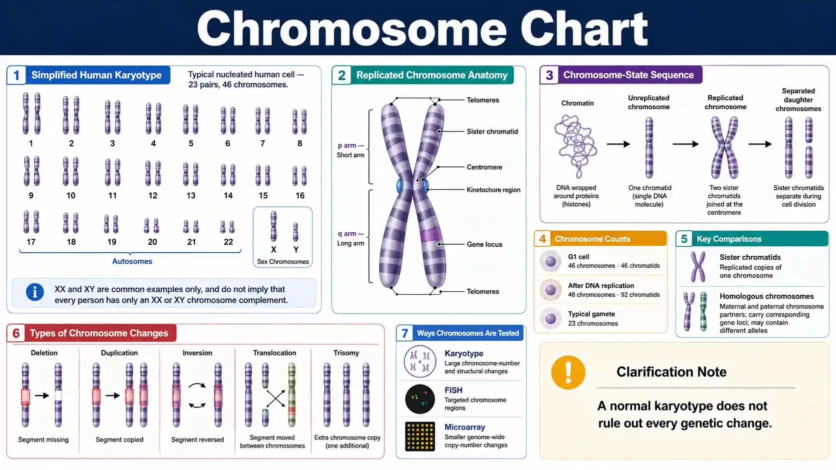
What is a chromosome?
A chromosome is one long DNA molecule packaged with histones and other proteins. Most nucleated human body cells contain 46 chromosomes arranged in 23 pairs.
The NHGRI chromosome fact sheet explains how chromosomes package DNA, protect chromosome ends, and segregate during cell division.
Typical diploid set
46 chromosomes
Most nucleated human body cells contain 23 chromosome pairs, with one homolog of each pair inherited from each biological parent.
Typical haploid set
23 chromosomes
Eggs and sperm normally contain one chromosome from each homologous pair after meiosis.
Replicated chromosome
2 sister chromatids
DNA replication creates two chromatids while the joined structure still counts as one chromosome.
Karyotype scope
Number and large structure
A karyotype can reveal large chromosome changes but does not read every gene or DNA base.
Essential chromosome questions
These direct answers establish chromosome terminology before the detailed charts.
How many chromosomes do humans usually have?
Most nucleated human body cells have 46 chromosomes arranged in 23 pairs.
How many chromosomes are in a human gamete?
A typical human egg or sperm contains 23 chromosomes.
What is a chromosome?
A chromosome is one DNA molecule packaged with histones and other proteins.
What is chromatin?
Chromatin is the DNA–protein material that forms chromosomes.
What is a chromatid?
A chromatid is one DNA copy within a replicated chromosome.
What are homologous chromosomes?
Homologous chromosomes are maternal and paternal partners that carry corresponding gene loci.
What does the centromere do?
The centromere supports kinetochore formation and accurate chromosome segregation.
What do telomeres do?
Telomeres protect the natural ends of linear chromosomes.
Does DNA replication double chromosome number?
No. DNA replication doubles chromatids and DNA content before sister chromatids separate.
What is a karyotype?
A karyotype is an ordered display or description of chromosome number and large-scale structure.
Can a normal karyotype rule out every genetic condition?
No. A normal karyotype cannot exclude many small or sequence-level genetic changes.
Is chromosome 22 the smallest human autosome?
No. Chromosome 21 is shorter than chromosome 22 in the GRCh38 reference assembly.
Chromosome Structure and Key Parts
A chromosome is one continuous DNA molecule packaged with proteins. Its visible form changes through the cell cycle, but the same core regions support replication, gene regulation, and segregation.
Swipe horizontally inside the table to view every column.
| Part or term | What it is | Primary role | Where it appears | Important distinction |
|---|---|---|---|---|
| Chromatin | DNA associated with histones and other proteins | Packages DNA and regulates access to genes | Throughout the nucleus between divisions | Chromatin is the material that forms chromosomes |
| Chromosome | One DNA molecule with its associated proteins | Carries genes and other functional DNA sequences | Present throughout the cell cycle | A duplicated chromosome still counts as one chromosome until sister chromatids separate |
| Sister chromatid | One of two nearly identical DNA copies after replication | Provides one copy for each daughter cell | Joined after S phase until separation | Two sister chromatids together form one replicated chromosome — Count by centromere |
| Centromere | Specialized chromosome region | Builds the kinetochore and supports accurate segregation — Segregation region | One functional centromere per normal linear chromosome | It is a region, not simply the narrowest drawing point |
| Kinetochore | Protein complex assembled on centromeric chromatin | Connects the chromosome to spindle microtubules | Forms during cell division | The kinetochore is protein machinery; the centromere is the chromosome region |
| p arm | The shorter arm extending from the centromere | Contains genes and chromosome bands | One side of a metaphase chromosome | p comes from the French word petit, meaning small |
| q arm | The longer arm extending from the centromere | Contains genes and chromosome bands | Opposite the p arm | q is the conventional label following p |
| Telomere | Specialized DNA-protein structure at each chromosome end | Protects chromosome ends and helps maintain stability — End protection | Two ends of each linear chromatid | Telomeres are not the same as centromeres |
| Replication origin | Site where DNA copying begins | Initiates DNA replication | Many origins along each human chromosome | Human chromosomes use many origins rather than one |
| Gene locus | Defined position of a gene or marker | Identifies where a sequence lies | Written by chromosome, arm, region, band, and sub-band | A locus is a position, not necessarily the entire chromosome |
Chromosome drawings are schematic and do not preserve true DNA length, band width, or three-dimensional shape.
- • A chromosome is not only the compact X-shaped form shown in many diagrams.
- • The X shape represents a replicated chromosome with two sister chromatids viewed during a condensed stage.
- • Each chromatid contains one continuous DNA double helix before chromosome ends and gaps are considered.
Download or export
Chromosome life cycle
Chromosomes change form without losing their identity
DNA remains organized into chromosomes throughout the cell cycle. Packaging, replication, condensation, attachment to the spindle, and separation change what a chromosome looks like and how many chromatids it contains.
Packaging
DNA → chromatin
Histones and other proteins compact long DNA molecules while preserving regulated access to genes.
Replication
1 → 2 chromatids
S phase copies one chromosome into two sister chromatids without immediately changing chromosome number.
Segregation
Centromere → kinetochore
The kinetochore links centromeric chromatin to spindle microtubules so chromosome copies separate accurately.
Inheritance
Homologs and gametes
Meiosis separates homologous chromosomes and then sister chromatids to produce haploid chromosome sets.
Count chromosomes by functional centromeres. Count DNA molecules or chromatids separately when comparing stages before and after replication.
Chromosome, chromatid, and homolog describe different relationships
The MedlinePlus chromosome overview shows the p arm, q arm, and centromere. Sister chromatids are replicated copies of one chromosome, while homologs are the maternal and paternal versions of the same chromosome number.
Human Chromosome Pairs and GRCh38 Reference Lengths
Human nuclear chromosomes include 22 numbered autosomes plus X and Y. Approximate lengths below use the GRCh38.p14 reference assembly and should not be treated as exact lengths for every individual genome.
Swipe horizontally inside the table to view every column.
| Chromosome | Type | Reference length | Approximate megabases | Usual copies in a diploid cell |
|---|---|---|---|---|
| 1 | Autosome | 248,956,422 bp — Largest numbered chromosome | 249.0 Mb | 2 |
| 2 | Autosome | 242,193,529 bp | 242.2 Mb | 2 |
| 3 | Autosome | 198,295,559 bp | 198.3 Mb | 2 |
| 4 | Autosome | 190,214,555 bp | 190.2 Mb | 2 |
| 5 | Autosome | 181,538,259 bp | 181.5 Mb | 2 |
| 6 | Autosome | 170,805,979 bp | 170.8 Mb | 2 |
| 7 | Autosome | 159,345,973 bp | 159.3 Mb | 2 |
| 8 | Autosome | 145,138,636 bp | 145.1 Mb | 2 |
| 9 | Autosome | 138,394,717 bp | 138.4 Mb | 2 |
| 10 | Autosome | 133,797,422 bp | 133.8 Mb | 2 |
| 11 | Autosome | 135,086,622 bp | 135.1 Mb | 2 |
| 12 | Autosome | 133,275,309 bp | 133.3 Mb | 2 |
| 13 | Autosome | 114,364,328 bp | 114.4 Mb | 2 |
| 14 | Autosome | 107,043,718 bp | 107.0 Mb | 2 |
| 15 | Autosome | 101,991,189 bp | 102.0 Mb | 2 |
| 16 | Autosome | 90,338,345 bp | 90.3 Mb | 2 |
| 17 | Autosome | 83,257,441 bp | 83.3 Mb | 2 |
| 18 | Autosome | 80,373,285 bp | 80.4 Mb | 2 |
| 19 | Autosome | 58,617,616 bp | 58.6 Mb | 2 |
| 20 | Autosome | 64,444,167 bp | 64.4 Mb | 2 |
| 21 | Autosome | 46,709,983 bp — Shortest numbered chromosome in GRCh38 | 46.7 Mb | 2 |
| 22 | Autosome | 50,818,468 bp | 50.8 Mb | 2 |
| X | Sex chromosome — Sex chromosome | 156,040,895 bp | 156.0 Mb | Usually 1 or 2 |
| Y | Sex chromosome — Sex chromosome | 57,227,415 bp | 57.2 Mb | Usually 0 or 1 |
bp = base pairs; Mb = million base pairs. Reference lengths include represented sequence and modeled gaps in GRCh38.p14.
- • Chromosome numbers were assigned roughly by apparent size, which is why chromosome 21 is shorter than chromosome 22 in modern sequence assemblies.
- • Reference assemblies are coordinate systems, not a complete description of every person’s chromosome sequence or structural variation.
- • Mitochondrial DNA is separate from the 46 nuclear chromosomes usually shown in a karyotype.
Download or export
Chromosome and Chromatid Counts Through Cell Division
Chromosome number is counted by functional centromeres. DNA replication doubles chromatids and DNA content without immediately doubling chromosome number.
Swipe horizontally inside the table to view every column.
| Cell state | Chromosomes per human cell | Chromatids or DNA molecules | Homologous pairs | What changed |
|---|---|---|---|---|
| Diploid cell in G1 | 46 | 46 | 23 pairs | Each chromosome has one chromatid |
| After S phase and in G2 | 46 — Chromosome number unchanged | 92 — DNA amount doubled | 23 replicated pairs | DNA replicated; sister chromatids remain joined |
| Metaphase of mitosis | 46 | 92 | 23 replicated pairs | Replicated chromosomes align individually |
| Anaphase of mitosis, within one dividing cell | 92 temporarily — Temporary count during separation | 92 | Homologs are not paired | Separated sister chromatids now count as daughter chromosomes |
| Each daughter cell after mitosis | 46 | 46 | 23 pairs | Each daughter receives one copy of every chromosome |
| Primary meiotic cell after S phase | 46 | 92 | 23 replicated homologous pairs | Homologs can pair and recombine |
| Each cell after meiosis I | 23 | 46 | No homologous pairs within one cell | Homologous chromosomes separated; sister chromatids remain joined |
| Each gamete after meiosis II | 23 — Haploid chromosome number | 23 | No homologous pairs | Sister chromatids separated |
Counts describe a typical human cell. Chromatid count is equivalent to the number of nuclear DNA molecules at these stages.
- • DNA content and chromosome number are different measurements.
- • A replicated X-shaped structure is one chromosome with two sister chromatids, not two chromosomes.
- • Mitochondrial DNA molecules are not included in these nuclear chromosome counts.
Download or export
Chromosome Set and Pairing Terminology
These terms describe chromosome sets, relationships, and inheritance. They should not be used interchangeably.
Swipe horizontally inside the table to view every column.
| Term | Definition | Human example | What is paired or counted | Common confusion |
|---|---|---|---|---|
| Diploid | Two chromosome sets | Most nucleated body cells have 46 chromosomes | One set from each biological parent | Diploid does not mean every chromosome is identical to its partner |
| Haploid | One chromosome set | Typical eggs and sperm have 23 chromosomes | One chromosome from each homologous pair | Haploid does not mean chromosomes are unreplicated |
| Homologous chromosomes | Maternal and paternal versions of the same numbered chromosome | The two chromosome 7 homologs | Similar gene loci in the same order | Homologs are not identical copies — Not identical copies |
| Sister chromatids | Replicated copies of one chromosome | Two chromatids joined after S phase | Nearly identical DNA copies from one original chromosome | Sisters are not the maternal and paternal homologs — Not homologs |
| Autosome | Any numbered non-sex chromosome | Chromosomes 1 through 22 | 22 pairs in a typical diploid cell | Autosome does not mean the chromosome lacks sex-related genes |
| Sex chromosome | X or Y chromosome | XX, XY, and other naturally occurring complements | Usually one chromosome pair or combination | Chromosome complement does not alone define every aspect of sex or gender |
| Karyotype | Ordered display or description of chromosomes | 46,XX or 46,XY are common examples | Number and large-scale structure | A karyotype is not a complete DNA sequence — Large-scale view |
| Genome | The complete set of DNA instructions | Nuclear genome plus mitochondrial genome | All chromosomes and organelle DNA | Genome is broader than karyotype |
| Allele | A sequence version at a locus | Different variants of a gene on homologs | Sequence variation at the same locus | An allele is not an entire chromosome |
Terminology describes biological organization; clinical notation may require more detailed cytogenetic conventions.
- • Homologous chromosomes contain corresponding loci but can carry different alleles and structural variants.
- • Sex-chromosome complements vary naturally and clinically beyond the simplified XX and XY examples.
- • Many mature human red blood cells have no nucleus and therefore no nuclear chromosome set.
Download or export
Browser-only educational tool
Compare chromosome structures and cell states
Select two concepts to compare DNA-molecule counts, centromeres, cell-cycle context, biological relationships, and identification clues. No information leaves the browser.
One chromosome after DNA replication
Replicated chromosome
- Category
- One chromosome after DNA replication
- DNA molecules
- 2
- Centromeres
- 1 functional joined centromere region before separation
- Usual context
- After S phase through metaphase
- Relationship
- Contains two sister chromatids joined by cohesion.
- Identification clue
- May appear X-shaped when highly condensed
Maternal and paternal chromosome partners
Homologous chromosome pair
- Category
- Maternal and paternal chromosome partners
- DNA molecules
- 2 before replication; 4 after replication
- Centromeres
- 2 before replication; still 2 replicated chromosomes after S phase
- Usual context
- Diploid cells; homologs pair directly during meiosis I
- Relationship
- Carry corresponding loci but can contain different alleles.
- Identification clue
- Two chromosomes of similar size and band pattern, not attached like sisters
Chromosome Number Changes
Chromosome-number changes can affect one chromosome or whole chromosome sets. Their effects depend on the chromosome, genes involved, timing, cell proportion, and biological context.
Swipe horizontally inside the table to view every column.
| Term | Meaning | Example notation or pattern | How it can arise | Interpretation limit |
|---|---|---|---|---|
| Aneuploidy | Gain or loss of one or more individual chromosomes — Individual chromosome change | 45 or 47 total chromosomes in a diploid lineage | Nondisjunction or chromosome loss | The term does not identify which chromosome changed |
| Monosomy | One copy of a particular chromosome instead of two | 45,X is one example | A chromosome is absent from a cell lineage | Viability and effects vary greatly by chromosome and mosaicism |
| Trisomy | Three copies of a particular chromosome | 47,+21 is one example | An extra chromosome is present | A count alone does not describe every clinical feature |
| Polyploidy | More than two complete chromosome sets | Triploidy has three sets | Whole-set segregation or fertilization errors | Common in plants but usually not compatible with typical human development |
| Mosaicism | Two or more cell populations with different chromosome complements | Some cells may be typical and others aneuploid | Change occurs after fertilization or during development | The tested tissue may not represent every tissue — Tissue-limited result |
| Nondisjunction | Failure of homologs or sister chromatids to separate normally | Can produce gametes with extra or missing chromosomes | Occurs in meiosis or mitosis | It is a mechanism, not a final diagnosis — Mechanism, not diagnosis |
| Uniparental disomy | Both homologs or chromosome copies derive from one parent | Chromosome count may still be 46 | Rescue of an earlier chromosome-number error or other mechanisms | A standard karyotype may not reveal parental origin |
Notation examples are educational summaries. Formal clinical reports use detailed ISCN cytogenetic nomenclature.
- • Aneuploidy can be constitutional, mosaic, or acquired in a tumor cell population.
- • A chromosome result must be interpreted with specimen type, test method, resolution, and clinical context.
- • Genetic counseling can help explain uncertain, prenatal, reproductive, or family implications.
Download or export
Chromosome Structural Changes and Rearrangements
Structural chromosome changes alter the amount, order, orientation, or location of chromosome material. Balanced appearance does not guarantee that no gene or regulatory region was disrupted.
Swipe horizontally inside the table to view every column.
| Change | What happens | Possible copy-number effect | Typical detection approach | Key limitation |
|---|---|---|---|---|
| Deletion | A chromosome segment is missing | Loss of DNA — DNA loss | Karyotype for large deletions; microarray or sequencing for smaller changes | Effect depends on size, genes, and mosaicism |
| Duplication | A chromosome segment is copied | Gain of DNA — DNA gain | Karyotype, microarray, or sequencing depending on size | Extra copies can have position and orientation effects |
| Inversion | A segment breaks and reinserts in reverse orientation | Often apparently balanced | Karyotype or sequencing | Breakpoints can disrupt genes; recombination can create unbalanced gametes |
| Reciprocal translocation | Segments are exchanged between nonhomologous chromosomes | May be balanced or unbalanced — Balance varies | Karyotype, FISH, or sequencing | Balanced carriers can have reproductive implications |
| Robertsonian translocation | Long arms of certain acrocentric chromosomes join | Can be balanced in a carrier | Karyotype | Chromosome count may be 45 without major net loss of essential long-arm material |
| Insertion | A segment moves into another location | May be balanced or unbalanced | Karyotype, FISH, microarray, or sequencing | Small or complex insertions can be difficult to resolve |
| Ring chromosome | Chromosome ends join to form a ring | Terminal material may be lost | Karyotype and complementary molecular testing | Rings can be unstable and mosaic |
| Isochromosome | One arm is duplicated while the other is lost | Simultaneous gain and loss | Karyotype and molecular confirmation | Clinical effect depends on the chromosome and mosaicism |
| Copy-number variant | DNA segment varies in copy number | Gain or loss | Chromosomal microarray or sequencing | Not every copy-number variant is harmful — May be benign or pathogenic |
Detection depends on change size, location, mosaic fraction, specimen, and laboratory platform.
- • A balanced rearrangement has no obvious large net gain or loss at the test resolution, but breakpoints may still matter.
- • A normal karyotype does not exclude small deletions, duplications, sequence variants, or low-level mosaicism.
- • Clinical laboratories classify findings using evidence, inheritance, population data, and phenotype information.
Download or export
Test resolution defines what a chromosome result can exclude
A karyotype examines chromosome number and large-scale structure. Microarray, targeted probes, and sequencing answer different questions. A normal result from one method does not guarantee that every smaller or different type of genetic change is absent.
Specimen matters
Blood, marrow, prenatal tissue, tumor, and skin samples can represent different cell populations.
Mosaicism matters
Low-level or tissue-limited mosaicism may be missed when few cells or one tissue are examined.
Clinical context matters
The same rearrangement can have different implications depending on inheritance, breakpoints, and phenotype.
Chromosome Tests and What They Can Detect
No single chromosome or genetic test detects every type of variation. The correct method depends on the clinical question and required resolution.
Swipe horizontally inside the table to view every column.
| Method | Best at detecting | Typical resolution | Common specimen context | Important limitation |
|---|---|---|---|---|
| Karyotype | Whole-chromosome gains, losses, and large rearrangements | Large cytogenetic changes | Blood, bone marrow, amniotic fluid, chorionic villi, or other dividing cells | Usually requires cultured or dividing cells and misses many small changes — Limited resolution |
| FISH | Targeted chromosome regions or known rearrangements | Targeted and higher resolution than routine karyotype | Interphase or metaphase cells | Only examines the probes selected — Targeted only |
| Chromosomal microarray | Genome-wide deletions and duplications | Smaller copy-number changes than karyotype — Genome-wide copy number | DNA from many specimen types | Usually does not identify balanced rearrangements or all sequence variants |
| QF-PCR or targeted aneuploidy assay | Rapid copy-number assessment for selected chromosomes | Targeted loci | Common in prenatal or confirmatory settings | Does not provide a full genome-wide chromosome survey |
| Genome or exome sequencing | Sequence variants and some structural variants | Base-level to larger events depending on pipeline | DNA from many specimen types | Coverage, repeat regions, mosaicism, and structural interpretation vary |
| Optical genome mapping | Large structural variants and complex rearrangements | High-molecular-weight DNA map | Specialized laboratory use | Availability and reporting standards vary |
Resolution is qualitative because platforms and laboratory validation thresholds differ.
- • A “normal” result means no reportable change was found by that specific method and resolution.
- • Testing decisions should be guided by the suspected condition, family history, specimen, urgency, and informed consent.
- • Unexpected, uncertain, or reproductive findings may require confirmatory testing and genetics consultation.
Download or export
A karyotype result needs the full laboratory interpretation
The MedlinePlus karyotype test guide explains that chromosome findings may be normal, abnormal, or require more testing. Discuss abnormal, uncertain, prenatal, reproductive, or potentially inherited findings with the ordering clinician or a genetics professional.
Basic Cytogenetic Notation Examples
Cytogenetic notation summarizes chromosome count and visible findings. These examples explain common components without replacing a formal laboratory report.
Swipe horizontally inside the table to view every column.
| Example | Plain-language meaning | Count component | Finding component | Caution |
|---|---|---|---|---|
| 46,XX | Forty-six chromosomes with two X chromosomes | 46 | XX | A common chromosome complement, not a complete health assessment |
| 46,XY | Forty-six chromosomes with one X and one Y chromosome | 46 | XY | A common chromosome complement, not a complete description of sex development |
| 47,XX,+21 | Forty-seven chromosomes, two X chromosomes, and an extra chromosome 21 | 47 | +21 — Extra chromosome | Clinical interpretation requires specimen and testing context |
| 45,X | Forty-five chromosomes with one X chromosome identified | 45 | X | Mosaic forms and structural X changes need separate notation |
| 46,XX,del(5p) | Forty-six chromosomes with a deletion involving the short arm of chromosome 5 | 46 | del(5p) — Structural deletion | A full report specifies breakpoints and method |
| 46,XY,t(9;22)(q34;q11.2) | A reciprocal translocation between chromosomes 9 and 22 at listed bands | 46 | t(9;22) | Acquired and constitutional contexts differ |
| mos 45,X/46,XX | Two detected cell lines with different chromosome complements | 45 and 46 | mos and slash-separated lines | Percentages depend on tested cells and tissue — Tissue sampling matters |
Examples use familiar simplified ISCN-style notation. Current formal ISCN rules contain additional detail and conventions.
- • The first number usually states the total chromosome count in the described cell line.
- • Symbols such as +, −, del, dup, inv, and t indicate gains, losses, deletions, duplications, inversions, and translocations.
- • Do not interpret a laboratory karyotype from notation alone without the report narrative and clinical context.
Download or export
Common Chromosome Mistakes and Corrections
These corrections resolve the most common errors in chromosome diagrams, counts, and genetic-test interpretation.
Swipe horizontally inside the table to view every column.
| Mistake | Correction | Why it matters | Better wording |
|---|---|---|---|
| Every chromosome is always X-shaped | The X shape appears when a replicated chromosome is highly condensed | Chromosomes exist throughout the cell cycle in less condensed forms | A metaphase chromosome often appears X-shaped |
| An X-shaped chromosome is two chromosomes | It is one replicated chromosome with two sister chromatids until they separate — Count by centromere | Chromosome number is counted by centromeres | One replicated chromosome contains two sister chromatids |
| Homologous chromosomes are identical | Homologs carry corresponding loci but can contain different alleles and sequence variants | Inheritance depends on maternal and paternal variation | Homologs are similar chromosome partners |
| DNA replication doubles chromosome number | Replication doubles DNA molecules and chromatids, not chromosome number before separation | DNA content and chromosome count must be kept separate | After S phase, 46 chromosomes contain 92 chromatids |
| Chromosome 22 is the smallest numbered chromosome | Chromosome 21 is shorter in current human reference assemblies | Historical numbering was based on early visual estimates | Chromosomes are numbered roughly, not perfectly, by size |
| A normal karyotype rules out every genetic condition | Karyotyping misses many small copy-number changes and sequence variants | Test resolution defines what can be excluded — Resolution limit | A normal karyotype excludes reportable large changes at that resolution |
| Balanced rearrangements never matter | Breakpoints and reproductive segregation can still have consequences | No visible net gain or loss does not guarantee no functional effect | Apparently balanced rearrangements need context |
| Every cell in one person has the same karyotype | Mosaicism, tumors, and specialized cells can differ | Specimen and tissue determine what was tested | Most constitutional cells usually share a chromosome complement |
| Sex chromosomes alone define gender | Chromosomes are one part of biological sex development and do not define gender identity — Use precise terminology | Genetics, development, anatomy, hormones, and identity are distinct concepts | Use precise language for chromosome complement |
| Chromosome size equals gene number | Chromosomes differ in gene density and repetitive DNA | Longer chromosomes do not always have proportionally more genes | Sequence length and gene content are separate attributes |
Corrections use standard introductory genetics language and acknowledge clinical and biological exceptions.
- • Simplified diagrams are useful for learning but should not be treated as literal microscopy images.
- • Clinical interpretation belongs in a validated laboratory report with professional context.
- • Reference genomes and cytogenetic nomenclature continue to improve as methods advance.
Download or export
Chromosome findings are not interpreted from a chart alone
Contact the ordering healthcare professional promptly when a report is marked critical or urgent, when prenatal results require time-sensitive decisions, or when a cancer cytogenetic report affects immediate treatment planning. The report, test method, specimen, and clinical history must be reviewed together.
Frequently asked questions
How many chromosomes do humans usually have?
Most nucleated human body cells have 46 chromosomes arranged as 23 pairs. Typical eggs and sperm have 23 chromosomes.
What is a chromosome made of?
A chromosome consists of one DNA molecule packaged with histone proteins and many other proteins that organize, regulate, copy, and repair the DNA.
What is the difference between chromatin and a chromosome?
Chromatin is DNA plus associated proteins. A chromosome is one organized unit of that chromatin containing one continuous nuclear DNA molecule.
What is the difference between a chromosome and a chromatid?
A chromatid is one DNA copy within a replicated chromosome. Two sister chromatids remain one chromosome until their centromeres separate.
Why does a chromosome look like an X?
The familiar X shape represents a duplicated, condensed chromosome with two sister chromatids joined at the centromere. Chromosomes are not always X-shaped.
What are homologous chromosomes?
Homologous chromosomes are the maternal and paternal versions of the same numbered chromosome. They carry corresponding loci but may have different alleles.
What is the centromere?
The centromere is a specialized chromosome region where the kinetochore assembles and spindle forces act during chromosome segregation.
What do telomeres do?
Telomeres protect the natural ends of linear chromosomes and help prevent chromosome ends from being treated like broken DNA.
What is a karyotype?
A karyotype is an ordered chromosome display or description used to assess chromosome number and large structural features.
Can a normal karyotype rule out every genetic disorder?
No. A normal karyotype does not exclude small deletions, duplications, sequence variants, low-level mosaicism, or changes outside its resolution.
What is trisomy?
Trisomy means that a cell contains three copies of a particular chromosome instead of the usual two in a diploid set.
What is monosomy?
Monosomy means that a cell contains one copy of a particular chromosome instead of the usual two in a diploid set.
What is a balanced translocation?
A balanced translocation rearranges chromosome material without an obvious large net gain or loss at the test resolution. Breakpoints and reproductive implications can still matter.
Why is chromosome 21 smaller than chromosome 22?
Early chromosome numbering relied on visual size estimates. Modern sequence assemblies show that chromosome 21 is shorter than chromosome 22.
Do all human cells contain 46 chromosomes?
No. Typical gametes contain 23 chromosomes, mature red blood cells lack a nucleus, and mosaic or acquired cell populations can have different complements.
When should chromosome results be discussed with a genetics professional?
Discuss results with a genetics professional when testing finds an abnormal, uncertain, mosaic, prenatal, reproductive, or potentially inherited chromosome change.
Sources
These references support the chromosome definitions, human chromosome counts, reference lengths, chromosome-change terminology, and testing limitations used throughout the chart.
National Human Genome Research Institute — Chromosomes Fact Sheet
https://www.genome.gov/about-genomics/fact-sheets/Chromosomes-Fact-Sheet
Explains chromosome number, chromosome structure, DNA packaging, centromeres, telomeres, and chromosome inheritance in humans.
National Human Genome Research Institute — Chromosome Genetics Glossary
https://www.genome.gov/genetics-glossary/Chromosome
Defines chromosomes and distinguishes the 22 human autosome pairs from the sex-chromosome pair.
MedlinePlus Genetics — What Is a Chromosome?
https://medlineplus.gov/genetics/understanding/basics/chromosome/
Describes DNA wrapped around histone proteins and labels the p arm, q arm, and centromere.
MedlinePlus Genetics — How Many Chromosomes Do People Have?
https://medlineplus.gov/genetics/understanding/basics/howmanychromosomes/
Explains the usual human complement of 23 chromosome pairs and introduces karyotypes.
MedlinePlus Genetics — Can Changes in Chromosome Number Affect Health?
https://medlineplus.gov/genetics/understanding/mutationsanddisorders/chromosomalconditions/
Explains aneuploidy, trisomy, monosomy, and how chromosome-number changes can arise.
MedlinePlus Genetics — Can Changes in Chromosome Structure Affect Health?
https://medlineplus.gov/genetics/understanding/mutationsanddisorders/structuralchanges/
Describes deletions, duplications, translocations, inversions, rings, and related chromosome rearrangements.
MedlinePlus — Karyotype Genetic Test
https://medlineplus.gov/lab-tests/karyotype-genetic-test/
Explains what a karyotype test examines and the limits of interpreting a normal or abnormal chromosome result.
NCBI Bookshelf — Chromosomes and Chromatin
https://www.ncbi.nlm.nih.gov/books/NBK9863/
Reviews chromatin organization, chromosome condensation, centromeres, telomeres, and chromosome behavior.
Genome Reference Consortium at NCBI — Human Genome Assembly GRCh38.p14
https://www.ncbi.nlm.nih.gov/grc/human/data
Provides reference chromosome lengths for the GRCh38.p14 human genome assembly.