Biology · Molecular Genetics
DNA Base Pair Chart for Pairing Rules, Strands, and Sequences
Compare A–T and C–G pairing, DNA and RNA bases, 5′ and 3′ direction, complements, reverse complements, replication, transcription, GC content, and sequence notation.
Sequence direction changes the answer. Label the input as DNA or RNA and state whether it is written 5′→3′ or 3′→5′ before calculating a complement or transcript.
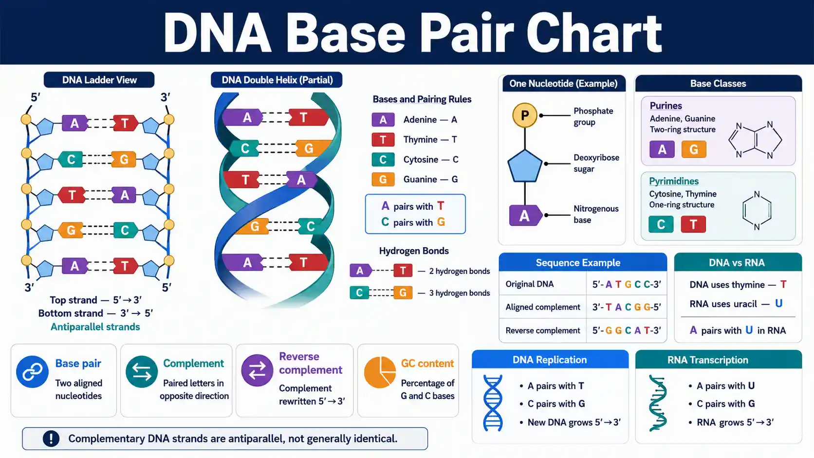
What pairs with each DNA base?
Adenine pairs with thymine, and cytosine pairs with guanine. The two DNA strands run in opposite chemical directions, so a directly aligned complement is antiparallel to the original sequence.
The NHGRI DNA fact sheet describes the sugar-phosphate backbones, opposite strand directions, and specific A–T and C–G pairing that form the double helix.
Canonical DNA pairs
A–T and C–G
Adenine pairs with thymine, while cytosine pairs with guanine in standard double-stranded DNA.
Strand orientation
Antiparallel
One DNA strand runs 5′→3′ while its directly aligned complementary partner runs 3′→5′.
RNA difference
U replaces T
RNA normally uses uracil in the pairing role occupied by thymine in DNA.
One base pair
2 nucleotides
A double-stranded base pair contains one nucleotide on each strand at the same paired position.
Essential DNA base-pair questions
These direct answers establish the core rules before the detailed sequence tables.
What are the DNA base-pairing rules?
Adenine pairs with thymine, and cytosine pairs with guanine.
What does A pair with in DNA?
Adenine pairs with thymine in canonical DNA.
What does C pair with in DNA?
Cytosine pairs with guanine in canonical DNA.
What does A pair with in RNA?
Adenine pairs with uracil in standard RNA base pairing.
How many hydrogen bonds join A and T?
The conventional Watson-Crick model shows two hydrogen bonds between A and T.
How many hydrogen bonds join G and C?
The conventional Watson-Crick model shows three hydrogen bonds between G and C.
What does antiparallel DNA mean?
Antiparallel DNA has one strand running 5′→3′ and the other running 3′→5′.
What is a reverse complement?
A reverse complement is the complementary sequence rewritten in the 5′→3′ direction.
Are complementary DNA strands identical?
No. Complementary strands use paired bases and opposite directions rather than identical letters.
What is a nucleotide?
A nucleotide contains a nitrogenous base, a five-carbon sugar, and phosphate.
What does GC content measure?
GC content measures the percentage of counted bases that are guanine or cytosine.
Does a base-pairing chart diagnose a genetic condition?
No. Base-pairing rules alone cannot establish variant meaning, sample identity, or clinical significance.
DNA Base Pairing Rules
Canonical Watson-Crick pairing joins one purine with one pyrimidine, helping maintain a consistent double-helix width and allowing either strand to specify its complement.
Swipe horizontally inside the table to view every column.
| Base on one DNA strand | Canonical partner | Base classes | Common hydrogen-bond model | Complement symbol |
|---|---|---|---|---|
| Adenine (A) | Thymine (T) | Purine–pyrimidine | 2 hydrogen bonds — Two-bond model | A ↔ T |
| Thymine (T) | Adenine (A) | Pyrimidine–purine | 2 hydrogen bonds | T ↔ A |
| Cytosine (C) | Guanine (G) | Pyrimidine–purine | 3 hydrogen bonds — Three-bond model | C ↔ G |
| Guanine (G) | Cytosine (C) | Purine–pyrimidine | 3 hydrogen bonds | G ↔ C |
Hydrogen-bond counts describe the conventional Watson-Crick base-pair model. DNA stability also depends strongly on base stacking, ionic conditions, sequence context, and temperature.
- • A base pair is two complementary nucleotides aligned across opposite nucleic-acid strands.
- • A–T and G–C pairing makes the two DNA strands complementary rather than identical.
- • Noncanonical pairs and mismatches can occur transiently or in specialized structures, but they are not the standard copying rule.
Download or export
From nucleotide to duplex
Base pairing connects chemistry, direction, and genetic information
A nucleotide contributes one base to a sequence. Complementary bases align across antiparallel backbones, while polymerases use the same pairing logic to copy DNA or synthesize RNA.
Pairing
A–T and C–G
Canonical purine-pyrimidine pairing lets either DNA strand specify the sequence of its partner.
Direction
Antiparallel strands
One backbone runs 5′→3′ while the directly aligned partner runs 3′→5′.
Copying
Template-directed synthesis
Polymerases read a template and extend a new nucleic-acid strand by adding nucleotides to its 3′ end.
Information
Sequence carries order
Base order stores biological information; pairing preserves and transfers that order during replication and transcription.
Always state the molecule type and strand direction before converting a sequence. A complement and a reverse complement contain the same paired bases in different written orientations.
Complementarity preserves sequence information without making the strands identical
The NCBI DNA structure chapter explains that complementary bases fit within an antiparallel double helix. Knowing one strand therefore determines the canonical sequence of the other strand.
DNA and RNA Bases, Sugars, and Pairing Roles
DNA and RNA share adenine, cytosine, and guanine. DNA normally uses thymine, while RNA normally uses uracil in the corresponding pairing role.
Swipe horizontally inside the table to view every column.
| Base | Abbreviation | Base class | Found in DNA | Found in RNA | Canonical pairing role |
|---|---|---|---|---|---|
| Adenine | A | Purine | Yes | Yes | Pairs with T in DNA or U in RNA |
| Guanine | G | Purine | Yes | Yes | Pairs with C |
| Cytosine | C | Pyrimidine | Yes | Yes | Pairs with G |
| Thymine | T | Pyrimidine | Yes | Usually no — DNA-specific standard base | Pairs with A in DNA |
| Uracil | U | Pyrimidine | Usually no — RNA-specific standard base | Yes | Pairs with A in RNA |
| Deoxyribose | — | Five-carbon sugar | Yes | No | Forms the DNA sugar-phosphate backbone |
| Ribose | — | Five-carbon sugar | No | Yes | Forms the RNA sugar-phosphate backbone |
A nucleotide contains a base, a five-carbon sugar, and one or more phosphate groups. A nucleoside contains the base and sugar without phosphate.
- • Purines have a two-ring structure; pyrimidines have a one-ring structure.
- • DNA can contain chemically modified bases, and some RNA molecules contain many modified nucleotides.
- • The table describes the standard bases used for introductory sequence notation.
Download or export
DNA Strand Direction and Complement Rules
DNA strands are antiparallel. When one strand is written 5-prime to 3-prime, its directly aligned partner runs 3-prime to 5-prime.
Swipe horizontally inside the table to view every column.
| Sequence task | Input strand | Required operation | Result orientation | Example |
|---|---|---|---|---|
| Aligned complement | 5′-ATGCC-3′ | Replace A↔T and C↔G without reversing | 3′ to 5′ — Antiparallel orientation | 3′-TACGG-5′ |
| Reverse complement | 5′-ATGCC-3′ | Complement, then reverse the character order | 5′ to 3′ — Standard sequence orientation | 5′-GGCAT-3′ |
| Coding-strand RNA sequence | 5′-ATGCC-3′ | Replace T with U | 5′ to 3′ | 5′-AUGCC-3′ |
| RNA from DNA template | 3′-TACGG-5′ | Use RNA complements A↔U and C↔G | 5′ to 3′ | 5′-AUGCC-3′ |
| Template from RNA | 5′-AUGCC-3′ | Find the antiparallel DNA complement | 3′ to 5′ | 3′-TACGG-5′ |
5′ and 3′ identify carbon positions in the sugar and therefore the chemical direction of a nucleic-acid strand.
- • A complement written directly beneath a 5′→3′ strand normally runs 3′→5′.
- • A reverse complement is rewritten in the conventional 5′→3′ direction.
- • Sequence software often reports both strands 5′→3′, which makes reverse-complement logic essential.
Download or export
Worked DNA Complement and Reverse-Complement Examples
These examples separate three operations that are often confused: direct complement, reverse complement, and RNA conversion.
Swipe horizontally inside the table to view every column.
| Original DNA, 5′→3′ | Aligned DNA complement, 3′→5′ | Reverse complement, 5′→3′ | Coding-like RNA, 5′→3′ | GC content |
|---|---|---|---|---|
| ATGC | TACG | GCAT | AUGC | 50% |
| GATTACA | CTAATGT | TGTAATC | GAUUACA | 28.6% |
| CCGGTA | GGCCAT | TACCGG | CCGGUA | 66.7% |
| AAAATTTT | TTTTAAAA | AAAATTTT — Self reverse-complementary sequence | AAAAUUUU | 0% |
| GCGCGC | CGCGCG | GCGCGC — Self reverse-complementary sequence | GCGCGC | 100% |
| ACGTN — Contains ambiguous base | TGCAN | NACGT | ACGUN | 50% of known bases |
GC content = (G + C) ÷ total counted A, T, G, and C bases × 100. N denotes an unknown or unspecified base and is excluded in the final example.
- • A sequence can equal its reverse complement; such sequence-level symmetry is called palindromic in molecular biology.
- • The coding-like RNA column is a letter substitution demonstration, not proof that the DNA segment is a transcribed coding strand.
- • Ambiguous IUPAC symbols require symbol-specific complement rules rather than simple A/T/G/C validation.
Download or export
Browser-only educational tool
Generate a DNA complement and reverse complement
Enter a short DNA sequence using A, T, G, and C. The tool labels strand direction, separates the direct complement from the reverse complement, and shows two common RNA conversions.
Spaces and line breaks are removed. Ambiguity symbols are intentionally rejected so every output remains deterministic.
- Input DNA
- 5′→3′ ATGCCGTA
- Aligned DNA complement
- 3′→5′ TACGGCAT
- Reverse complement
- 5′→3′ TACGGCAT
- Coding-like RNA
- 5′→3′ AUGCCGUA
- RNA transcribed from the input as template
- 5′→3′ UACGGCAU
- Composition
- 8 nt · 4 G/C bases · 50.0% GC
Base Pairing in DNA Replication and Transcription
Both processes use a template strand, but they produce different molecules and use different enzymes and base sets.
Swipe horizontally inside the table to view every column.
| Feature | DNA replication | Transcription | Pairing rule | Key distinction |
|---|---|---|---|---|
| Main product | Double-stranded DNA | RNA | Template-directed complementarity | Replication copies DNA; transcription makes an RNA product |
| Template use | Both parental strands serve as templates | One DNA strand is used for a given transcript | Read by polymerase | Genes can use different template strands |
| New-strand bases | A, T, G, C | A, U, G, C — Uracil replaces thymine | A pairs with T or U; G pairs with C | RNA uses U instead of T |
| Synthesis direction | 5′→3′ | 5′→3′ | Nucleotides add to the 3′ end — Polymerase direction rule | Template is read 3′→5′ |
| Primer requirement | DNA polymerases generally require a primer | RNA polymerase initiates without a conventional primer | Initial base pairing stabilizes synthesis | Initiation mechanisms differ |
| Extent | Genome or replicating DNA molecule | Selected transcription unit | Local complementarity | Transcription does not copy the whole genome at once |
| Accuracy systems | High-fidelity copying and repair pathways | Proofreading and RNA quality-control vary | Mismatches can be recognized | Error consequences and correction differ |
This is a broad educational comparison. Polymerase families, organisms, organelles, and specialized pathways add important exceptions.
- • During replication, each parental strand guides synthesis of a new complementary DNA strand.
- • During transcription, RNA polymerase uses the DNA template strand to synthesize RNA 5′→3′.
- • The RNA sequence usually matches the DNA coding strand except that U replaces T.
Download or export
RNA polymerase reads the DNA template strand
The OpenStax transcription guide describes RNA synthesis in the 5′→3′ direction. The transcript is complementary to the template strand and usually matches the coding strand except that U replaces T.
DNA Sequence Size and Composition Units
Sequence length and composition are reported with different units depending on whether the molecule is double-stranded, single-stranded, or summarized at genome scale.
Swipe horizontally inside the table to view every column.
| Term or unit | Meaning | Typical use | Example | Interpretation caution |
|---|---|---|---|---|
| Base pair | One paired position in double-stranded nucleic acid | Double-stranded DNA length | 500 bp DNA fragment | One base pair contains two nucleotides — Two nucleotides per pair |
| Nucleotide | One base-sugar-phosphate unit | Single-stranded DNA or RNA length | 100 nt RNA | nt and bp are not interchangeable without strand context |
| Kilobase pair | 1,000 base pairs | Genes, PCR products, plasmid features | 2.4 kb | Often shortened informally to kb |
| Megabase pair | 1,000,000 base pairs | Chromosomes and large genomic regions | 50 Mb | Reference assemblies can include gaps or alternate sequence |
| Gigabase pair | 1,000,000,000 base pairs | Whole-genome scale | A 3 Gb-scale haploid genome assembly | Personal genomes and assemblies differ |
| GC content | Fraction of counted bases that are G or C | Sequence composition and assay design | 42% GC | Does not alone predict exact melting temperature — Composition is not temperature |
| Read length | Number of sequenced bases in one read | Sequencing platforms | 150 nt read | Quality can vary across a read |
| Coverage or depth | Average number of reads overlapping a position | Sequencing confidence | 30× mean depth | Average depth does not guarantee every base has 30 reads |
Decimal prefixes are standard in sequence reporting: 1 kb = 10³ bp, 1 Mb = 10⁶ bp, and 1 Gb = 10⁹ bp.
- • A 100 bp double-stranded DNA fragment contains 100 paired positions and 200 nucleotides total.
- • GC percentage should state how ambiguous or missing bases were handled.
- • Sequence length, mapped length, assembled length, and biological molecule length can differ.
Download or export
Hydrogen-bond counts do not fully predict DNA melting behavior
GC content can influence duplex stability, but exact melting temperature also depends on sequence order, fragment length, neighboring bases, salt, magnesium, strand concentration, mismatches, and assay conditions. Use a validated thermodynamic model for primer or probe design.
Sequence context matters
Two sequences with the same GC percentage can melt at different temperatures.
Solution conditions matter
Ions and strand concentration affect duplex formation and stability.
Mismatches matter
Mismatch type and position can change binding in ways that simple bond counting misses.
DNA Mismatches and Sequence-Change Terms
A mismatch describes incompatible paired bases in a duplex. A variant describes a sequence difference relative to another sequence or reference and may or may not affect function.
Swipe horizontally inside the table to view every column.
| Term | What changes | Example | Possible consequence | Important distinction |
|---|---|---|---|---|
| Mismatch | Opposing bases do not form the intended canonical pair | A opposite C | Polymerase stalling, repair, or mutation if unresolved | A mismatch is a molecular pairing state, not automatically an inherited variant — Pairing state, not diagnosis |
| Substitution | One nucleotide is replaced by another | A→G | May be silent, regulatory, missense, nonsense, or neutral | Does not change sequence length |
| Transition | Purine replaces purine or pyrimidine replaces pyrimidine | A↔G or C↔T | Context-dependent | A subtype of substitution |
| Transversion | Purine and pyrimidine replace one another | A↔C, A↔T, G↔C, or G↔T | Context-dependent | A subtype of substitution |
| Insertion | One or more nucleotides are added | ACG→ACTG | Can alter spacing or reading frame | Sequence length increases |
| Deletion | One or more nucleotides are removed | ACGT→AGT | Can remove a motif or alter reading frame | Sequence length decreases |
| Indel | Insertion or deletion described as one variant class | Small sequence gain or loss | Context-dependent | The term does not specify functional impact |
| Single-nucleotide variant | One genomic position differs from a reference or comparison | Reference C, sample T | May be benign, uncertain, or clinically relevant | Variant is not synonymous with disease-causing mutation — Neutral term |
| Ambiguous base call | The exact base is uncertain or mixed | N in a consensus sequence | Requires quality review or additional data | Not necessarily a biological variation — May be technical uncertainty |
Variant interpretation depends on genomic position, sequence context, evidence, inheritance, assay quality, and the reference sequence used.
- • DNA repair systems can correct many mismatches before they become fixed sequence changes.
- • A sequence difference can be common and harmless, rare and harmless, uncertain, or functionally important.
- • Clinical interpretation should not rely on base-pair identity alone.
Download or export
DNA Sequence Notation and IUPAC Ambiguity Codes
Sequence notation should state direction, molecule type, and whether symbols represent exact bases or a set of possible bases.
Swipe horizontally inside the table to view every column.
| Symbol or notation | Meaning | DNA complement | Example use | Caution |
|---|---|---|---|---|
| 5′ and 3′ | Chemical strand ends and reading direction | Opposite strand runs antiparallel | 5′-ACGT-3′ | Do not omit direction when orientation matters |
| N | Any base: A, C, G, or T | N | ACNGT | Can indicate unknown sequence or an intentional wildcard |
| R | Purine: A or G | Y — R complements Y | R-rich motif | Complement changes to the opposite ambiguity class |
| Y | Pyrimidine: C or T | R | Y-rich motif | Not the amino-acid symbol context |
| W | A or T | W | Weak-pair class | Name reflects the conventional two-hydrogen-bond model |
| S | C or G | S | Strong-pair class | Name reflects the conventional three-hydrogen-bond model |
| K | G or T | M — K complements M | Degenerate primer position | Use the correct reverse-complement symbol |
| M | A or C | K | Degenerate primer position | Use the correct reverse-complement symbol |
| B | C, G, or T; not A | V | Consensus sequence | Represents three possible bases |
| D | A, G, or T; not C | H | Consensus sequence | Represents three possible bases |
| H | A, C, or T; not G | D | Consensus sequence | Represents three possible bases |
| V | A, C, or G; not T | B | Consensus sequence | Represents three possible bases |
IUPAC ambiguity symbols describe sets of possible bases. They do not assign probabilities to those possibilities.
- • Uppercase and lowercase sequence letters usually carry the same base identity unless a software format assigns special meaning.
- • Reverse-complement calculations with ambiguity codes must complement each symbol and reverse the order.
- • Some file formats use additional symbols or quality fields; always follow the relevant format specification.
Download or export
Common DNA Base-Pairing Mistakes
Most base-pairing errors come from mixing strand orientation, DNA versus RNA alphabets, or molecular terminology.
Swipe horizontally inside the table to view every column.
| Mistake | Why it is incorrect | Correct approach | Quick check | Why it matters |
|---|---|---|---|---|
| Writing both aligned strands 5′→3′ | Aligned DNA strands are antiparallel | Write the partner 3′→5′ or explicitly use a reverse complement — Label direction | Check the end labels | Orientation affects primers, transcripts, and sequence matching |
| Calling a complement a reverse complement | Complementing and reversing are separate operations | Name the output according to its orientation | Compare TACG with GCAT for ATGC | Software searches often require the reverse complement |
| Using U in a DNA sequence | Standard DNA notation uses T | Use U for RNA and T for DNA | Check molecule type | Mixing alphabets can invalidate analysis |
| Using T in an RNA transcript | Standard RNA notation uses U | Replace coding-strand T with U | Scan for T characters | Transcript and codon interpretation changes |
| Counting one base pair as one nucleotide total | A double-stranded base pair contains two nucleotides | State bp for paired positions and nt for nucleotides | 100 bp = 200 nucleotides total | Length comparisons become inconsistent |
| Assuming three hydrogen bonds make every GC-rich DNA exactly three-halves as stable | Duplex stability is sequence- and condition-dependent | Use an appropriate melting-temperature model | Include salt, length, and sequence context | Simplified bond counting cannot replace thermodynamics — Thermodynamic oversimplification |
| Treating the coding strand as the strand read by RNA polymerase | RNA polymerase reads the template strand | Use the coding strand only as the RNA-like reference sequence | RNA matches coding DNA except U for T | Strand-role errors reverse the transcript |
| Assuming every sequence difference causes disease | Many variants are neutral or uncertain | Interpret variants with evidence and context | Separate sequence identity from clinical meaning | Prevents unsupported conclusions — No clinical inference |
| Ignoring ambiguous symbols | N, R, Y, and other codes can represent multiple bases | Use IUPAC-aware validation and complement rules | Do not silently delete symbols | Ambiguity affects matching and assay design |
These checks support education and sequence handling. Laboratory and clinical workflows require validated software, controls, and professional interpretation.
- • Always label molecule type and strand direction before converting a sequence.
- • For long or clinically important sequences, use validated tools and retain the original record and reference coordinates.
- • Do not infer gene function, pathogenicity, or ancestry from a short base-pairing exercise.
Download or export
A short sequence conversion cannot establish biological or clinical meaning
Complementing a sequence proves only the expected pairing operation for the supplied letters. It does not verify sample quality, genome position, gene identity, reading frame, organism, inheritance, pathogenicity, or personal identity.
Frequently asked questions
What are the DNA base-pairing rules?
Adenine pairs with thymine, and cytosine pairs with guanine in canonical double-stranded DNA.
How many hydrogen bonds join A and T?
The conventional Watson-Crick model shows two hydrogen bonds between adenine and thymine.
How many hydrogen bonds join G and C?
The conventional Watson-Crick model shows three hydrogen bonds between guanine and cytosine.
What is a DNA base pair?
A DNA base pair is two complementary nucleotides aligned across opposite strands of a DNA duplex.
Why are DNA strands called antiparallel?
DNA strands are antiparallel because one runs 5′→3′ while the aligned partner runs 3′→5′.
What is the complement of 5′-ATGC-3′?
The directly aligned complement is 3′-TACG-5′.
What is the reverse complement of 5′-ATGC-3′?
The reverse complement is 5′-GCAT-3′.
What replaces thymine in RNA?
Uracil replaces thymine in standard RNA, so adenine pairs with uracil during RNA base pairing.
Are the two DNA strands identical?
No. The two strands are complementary and antiparallel, not generally identical.
What is the difference between a base and a nucleotide?
A base is the nitrogen-containing component, while a nucleotide contains a base, sugar, and phosphate group.
Does one base pair equal one nucleotide?
No. One double-stranded base pair contains two nucleotides, one on each strand.
What does GC content mean?
GC content is the percentage of counted bases in a sequence that are guanine or cytosine.
Does higher GC content always mean a specific melting temperature?
No. GC content influences duplex stability, but exact melting temperature also depends on sequence, length, salt, concentration, and assay conditions.
Which DNA strand matches an RNA transcript?
The RNA sequence matches the DNA coding strand except that RNA uses U instead of T; it is complementary to the DNA template strand.
What does N mean in a DNA sequence?
N means that the base is unknown, unspecified, or allowed to be any of A, C, G, or T.
Can a short DNA sequence identify a disease or person?
Not reliably. Interpretation requires validated methods, reference context, sequence quality, and often much more genomic and clinical information.
Sources
These references support the DNA and RNA base definitions, pairing rules, antiparallel strand orientation, replication, transcription, and sequence terminology used throughout the chart.
National Human Genome Research Institute — Base Pair
https://www.genome.gov/genetics-glossary/Base-Pair
Defines a base pair and states the canonical DNA pairing rules: adenine with thymine and cytosine with guanine.
National Human Genome Research Institute — Deoxyribonucleic Acid (DNA) Fact Sheet
https://www.genome.gov/about-genomics/fact-sheets/Deoxyribonucleic-Acid-Fact-Sheet
Explains the double helix, sugar-phosphate backbones, opposite strand directions, and specific complementary base pairing.
National Human Genome Research Institute — Nucleotide
https://www.genome.gov/genetics-glossary/Nucleotide
Defines nucleotides as the sugar, phosphate, and nitrogenous-base building blocks of DNA and RNA.
NCBI Bookshelf — The Structure and Function of DNA
https://www.ncbi.nlm.nih.gov/books/NBK26821/
Describes complementary, antiparallel DNA strands and the structural consequences of purine-pyrimidine pairing.
NCBI Bookshelf — DNA Replication Mechanisms
https://www.ncbi.nlm.nih.gov/books/NBK26850/
Reviews template-directed DNA synthesis, complementary nucleotides, replication forks, primers, and polymerase directionality.
OpenStax — Nucleic Acids
https://openstax.org/books/biology-2e/pages/3-5-nucleic-acids
Compares DNA and RNA bases, purines and pyrimidines, nucleotide structure, and uracil pairing in RNA.
OpenStax — The Structure of DNA
https://openstax.org/books/concepts-biology/pages/9-1-the-structure-of-dna
Illustrates the double helix, A-T and G-C pairing, and the commonly taught two-versus-three hydrogen-bond model.
OpenStax — Prokaryotic Transcription
https://openstax.org/books/biology-2e/pages/15-2-prokaryotic-transcription
Explains template-strand reading, RNA synthesis in the 5-prime to 3-prime direction, and replacement of thymine with uracil in RNA.
NCBI GenBank — FASTA Format for Nucleotide Sequences
https://www.ncbi.nlm.nih.gov/genbank/fastaformat
Provides current NCBI sequence-format guidance and requires IUPAC nucleotide symbols, including N for ambiguous bases.