Health & Medical · Iris color and genetics reference
Eye Color Chart: Iris Shades, Genetics and Health
Compare common iris shades, understand melanin and polygenic inheritance, follow baby eye-color development, recognize heterochromia, and separate ordinary lighting effects from changes that need an eye examination.
An eye-color label is descriptive, not diagnostic. Seek professional care for a new one-sided change, pain, redness, light sensitivity, trauma, an unusual pupil, or reduced vision. Read the ChartsLoom Disclaimer.
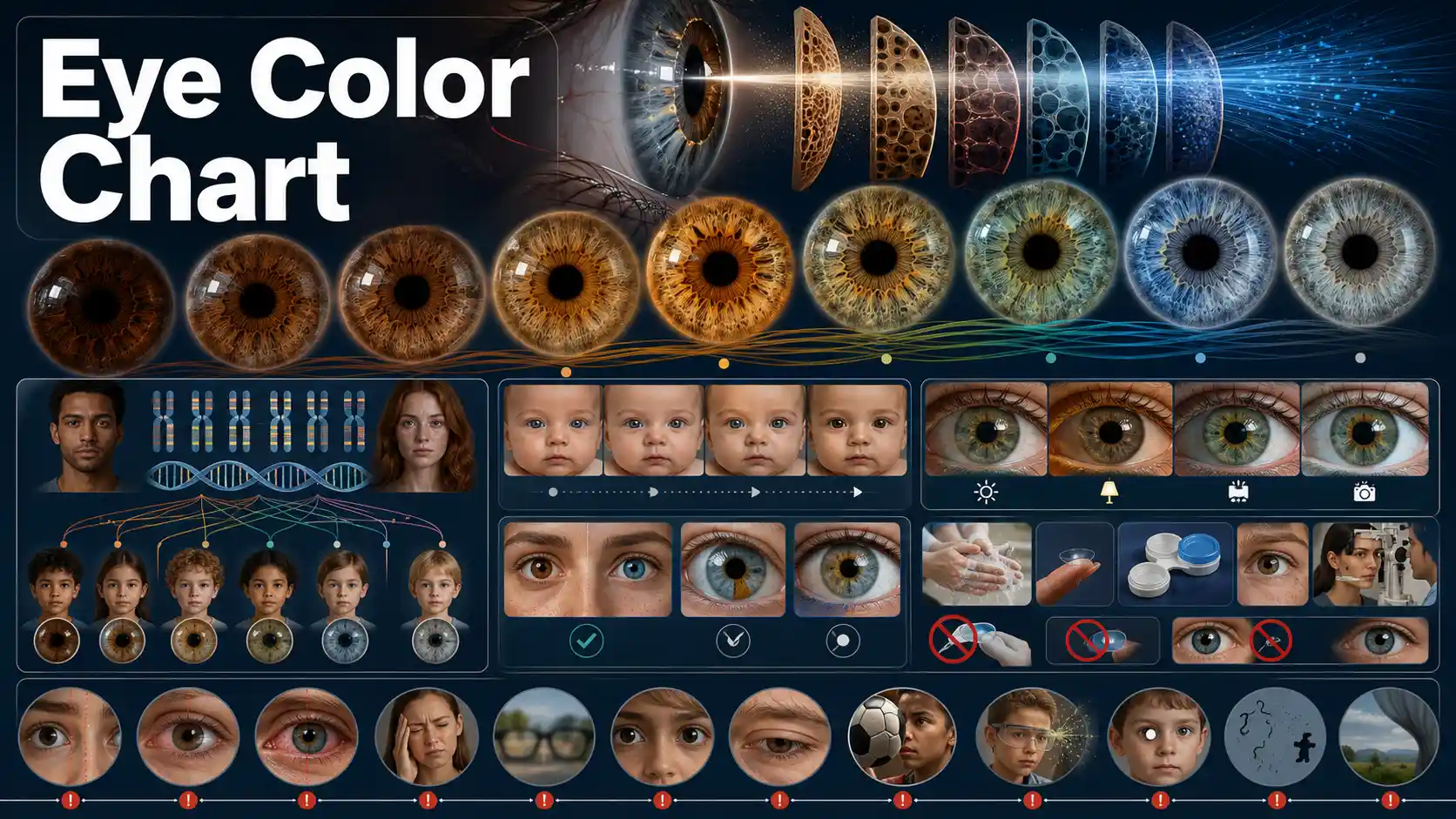
What determines whether eyes look brown, hazel, green, blue or gray?
Eye color reflects the amount and distribution of melanin in the iris, iris tissue structure, and the way light scatters. Many genes contribute, so natural shades form a continuum rather than a simple dominant-versus-recessive grid.
MedlinePlus explains the polygenic basis of eye color, including the major roles of OCA2 and HERC2.
Main pigment
Melanin
Amount and distribution strongly influence visible shade.
Inheritance
Polygenic
Many genes contribute; one dominant gene is not enough.
Color labels
Overlap
Hazel, amber, green, blue and gray form a continuum.
New change
Check it
One-sided or symptomatic changes need professional review.
Common Eye Color Shades and Iris Patterns
Every iris is unique. These broad categories describe visible color, not a diagnostic classification or a precise melanin measurement.
Swipe horizontally inside the table to view every column.
| Broad category | Typical appearance | Pigment and optical context | Common variations | Important limitation |
|---|---|---|---|---|
| Dark brown | Deep brown that may look nearly black in low light | Higher visible iris pigmentation | Warm, neutral, or reddish-brown undertones | The pupil and iris can blend together in dim photographs |
| Medium brown | Clearly brown with visible radial texture | Substantial melanin with more visible stromal detail | Golden flecks, darker limbal ring, lighter central zone | Lighting can shift the apparent warmth |
| Light brown | Honey, caramel, or light chestnut appearance | Moderate pigment with stronger light reflection | Amber-brown, golden-brown, mixed brown-green | May be confused with hazel or amber |
| Amber | Relatively uniform golden, copper, or yellow-brown tone | Warm pigment appearance with less obvious green mixing | Copper, honey, russet, golden | No universal clinical boundary separates amber from light brown |
| Hazel | Mixed brown, green, and gold that can vary across the iris | Uneven pigment distribution and optical scattering | Brown center with green outer iris, gold flecks, sector-like blends | Hazel is a descriptive category, not one fixed color |
| Green | Green or green-gray appearance, often with gold or brown flecks | Lower-to-moderate visible pigment plus light scattering | Olive, moss, sea-green, gray-green | Camera processing can exaggerate saturation |
| Blue | Blue, blue-gray, or blue-green appearance | Lower visible anterior iris pigment with light scattering | Pale blue, slate blue, blue-green, dark limbal ring | Blue color is not created by blue pigment |
| Gray | Silver, slate, blue-gray, or green-gray appearance | Low visible pigment with stromal structure and light scattering | Steel gray, pale gray, gray-blue, gray-green | Gray and blue overlap on a continuum |
- • Iris color exists on a continuum; labels differ among observers, cultures, cameras, and lighting conditions.
- • Population frequencies vary substantially by geography and ancestry, so a chart should not assign identity from appearance.
- • Color categories do not predict visual acuity, personality, ancestry, or eye health by themselves.
Download or export
Eye color is a spectrum, not seven separate boxes
The same iris can contain a dark limbal ring, a lighter outer field, a brown central ring, radial streaks, and scattered flecks. Broad labels help communication but cannot capture every pattern.
Dark brown
Higher visible pigment
Brown
Substantial pigment
Amber
Warm golden appearance
Hazel
Mixed distribution
Green
Pigment + scattering
Blue
Lower visible pigment
Gray
Low pigment + structure
What Creates the Appearance of Eye Color?
Visible iris color reflects pigment amount and distribution together with the way light interacts with iris tissue.
Swipe horizontally inside the table to view every column.
| Factor | How it affects appearance | Can it change the observed color? | Practical interpretation |
|---|---|---|---|
| Melanin amount | More visible iris melanin generally produces darker brown appearance | Yes, especially during early development | Melanin level is a major contributor but not the only contributor |
| Pigment distribution | Uneven pigment creates flecks, rings, sectors, and mixed colors | Usually stable, but true changes need context | Hazel and heterochromia patterns often reflect distribution |
| Iris stroma | Microscopic tissue structure changes how shorter wavelengths scatter | Usually stable | Helps explain blue, gray, and green appearances without blue or green pigment |
| Limbal ring | A darker outer border increases apparent contrast | Can look stronger or weaker with age and lighting | Does not change the underlying iris category |
| Pupil size | A larger or smaller pupil exposes a different amount of iris and changes contrast | Yes, temporarily | Bright and dim settings can make the same iris look different |
| Ambient lighting | Warm, cool, direct, or diffuse light changes reflected color | Yes, temporarily | Compare colors in indirect daylight for consistency |
| Camera and screen | White balance, exposure, filters, and display calibration alter color | Yes, in photos and video | A photograph is not a clinical color measurement |
| Clothing and background | Nearby colors change perceived contrast | Yes, visually | This is an optical context effect, not pigment change |
| Contact lenses | Tinted lenses cover or blend with natural iris color | Yes, while worn | Decorative lenses require professional fitting and a prescription |
| Eye disease, injury, surgery, or medicine | Some conditions or treatments can alter pigment or iris structure | Yes, sometimes permanently | New or one-sided change should be examined |
- • Apparent color change is common when light, pupil size, camera settings, or surroundings change.
- • True pigment change is less common and should be distinguished from reflections, redness, corneal clouding, or a change in pupil appearance.
Download or export
Eye Color Genetics: What Is Known and What Is Not
Eye color is polygenic. A simple brown-dominant and blue-recessive school model cannot reliably predict every child.
Swipe horizontally inside the table to view every column.
| Genetic concept | Role in eye color | What it can explain | What it cannot guarantee |
|---|---|---|---|
| OCA2 | Influences production and handling of melanin in the iris | A major share of brown-to-blue variation | A single final color by itself |
| HERC2 | Regulates OCA2 activity through a nearby control region | Strong association with lighter versus darker irises | All green, hazel, gray, or amber variation |
| Other pigmentation genes | Multiple genes make smaller contributions | Continuous shade and pattern differences | A simple two-gene prediction |
| Polygenic inheritance | Many variants combine to influence pigment and structure | Why siblings can have different eye colors | A certain child outcome from parental labels alone |
| Ancestry and population history | Changes the frequency of variants in populations | Why color frequencies differ by region | An individual’s identity or exact ancestry |
| Genetic recombination | Each child receives a different combination of parental variants | Unexpected-looking family combinations | The same result for every sibling |
| Mosaicism and developmental variation | Different cell populations may carry or express pigment-related differences | Some sectoral or asymmetric patterns | Whether a pattern is harmless without examination |
| Albinism-related genes | Can markedly reduce pigment in the iris and other tissues | Very light irises and associated visual-development concerns | Diagnosis based on eye color alone |
- • Parental eye-color charts provide rough possibilities, not Mendelian guarantees.
- • Genetic testing for a medical condition is different from consumer prediction of cosmetic traits.
- • Eye color alone cannot establish biological parentage or ancestry.
Download or export
Parental eye-color charts show possibilities, not guarantees
Siblings inherit different combinations of variants. A consumer prediction grid cannot include every pigmentation gene, regulatory variant, ancestry pattern, or developmental effect.
Baby Eye Color Development Timeline
Some infants are born with lighter-looking irises that darken as pigment develops. Timing varies widely and is not identical across ancestries or children.
Swipe horizontally inside the table to view every column.
| Stage | What may be observed | What is usually normal | When to ask a clinician |
|---|---|---|---|
| At birth | Iris may appear blue-gray, gray, brown, or another shade | Color may be difficult to judge under newborn lighting | Unusual pupil shape, cloudiness, absent red reflex, or obvious structural difference |
| First 3 months | Color can remain similar or begin to darken | Gradual bilateral change without symptoms | Redness, swelling, light sensitivity, discharge, or poor visual behavior |
| 3–6 months | Brown pigment may become more noticeable in some babies | Increasing warmth, flecks, or central darkening | One eye changing differently with eyelid droop or unequal pupils |
| 6–12 months | Many infants approach a more stable visible color | Subtle continued development can occur | Sudden change, pain, trauma, or vision concern |
| Second and third years | Some children continue subtle shade or pattern changes | Slow, symmetric maturation without symptoms | New heterochromia, progressive asymmetry, or a white pupil in photographs |
| Later childhood | Color is generally stable | Minor apparent changes with light and pupil size | True new pigment loss, darkening, inflammation, or visual symptoms |
- • A baby’s final color cannot be determined accurately from a newborn photograph.
- • Do not delay routine pediatric vision screening while waiting for eye color to settle.
- • A white pupil reflex is not an eye-color variant and requires prompt medical assessment.
Download or export
Baby eye color can develop gradually during the first year
Some lighter-looking newborn irises darken as pigmentation develops, while many babies with brown eyes at birth retain brown eyes. Read the AAP newborn-eye overview.
Eye color pattern and change checker
Select a broad shade, pattern, timing, and symptoms. This browser-only tool provides context; it does not analyze photographs, measure pigment, predict genetics, or diagnose eye disease.
This is a descriptive color category, not a medical result
Hazel appearance with mostly uniform color.
Use the same indirect daylight and camera settings for comparisons. Natural color labels overlap and cannot measure iris melanin precisely.
Heterochromia Types and Evaluation Context
Heterochromia describes different iris colors between eyes or within one iris. Long-standing patterns are often benign, while acquired change deserves assessment.
Swipe horizontally inside the table to view every column.
| Pattern | Appearance | Possible context | Recommended approach |
|---|---|---|---|
| Complete heterochromia | One iris is a different overall color from the other | Congenital variation, genetic condition, injury, inflammation, surgery, or medicine | Document onset; examine if new, progressive, or accompanied by symptoms |
| Sectoral heterochromia | A wedge-shaped section differs from the rest of the iris | Developmental pigment variation or acquired change | Long-standing stable pattern may be benign; new sector needs examination |
| Central heterochromia | A ring around the pupil differs from the outer iris | Common pigment-distribution pattern | Usually descriptive rather than dangerous when stable and symptom-free |
| Subtle bilateral variation | Both eyes share similar mixed rings, flecks, or gradients | Normal iris texture and uneven pigment distribution | Compare under the same lighting before calling it a change |
| Acquired heterochromia | A new difference develops after previously similar color | Eye inflammation, trauma, surgery, medicine, nerve-related disorder, or pigment change | Arrange an eye examination |
| Heterochromia with eyelid droop or unequal pupils | Color difference plus asymmetry of pupil or lid | Can occur with nerve-pathway conditions | Needs clinical assessment, especially in a child or when newly developed |
| Heterochromia with pain, redness, or blurred vision | Color difference plus active symptoms | Inflammation, injury, infection, or other eye disease | Seek prompt or urgent eye care |
- • Heterochromia itself does not reveal the cause.
- • Congenital patterns should still be mentioned during routine eye care, particularly when other eye, skin, hair, hearing, or neurologic differences are present.
Download or export
Heterochromia describes a pattern, not its cause
A stable difference present from birth is often benign, but acquired change may follow inflammation, injury, surgery, medicine, or another condition. Review the AAO heterochromia guide.
Apparent Eye Color Change Versus True Iris Change
Many day-to-day differences are optical. New pigment or structural change, especially in one eye, requires a different response.
Swipe horizontally inside the table to view every column.
| Observation | More likely explanation | Simple check | When to seek care |
|---|---|---|---|
| Both eyes look lighter outdoors | Brighter light, smaller pupils, and stronger iris detail | Compare in the same indirect daylight | Routine care unless symptoms or true asymmetry appear |
| Eyes look greener with certain clothing | Color contrast from surroundings | Change background and remove filters | No urgent action if both eyes are unchanged |
| Photos show different colors on different phones | White balance, exposure, flash, and screen calibration | Use the same camera and lighting | Do not diagnose from images alone |
| Color changes after putting in tinted contacts | Lens color or poor lens fit | Remove lenses and inspect the natural iris | Stop wear and seek care for pain, redness, discharge, or blurred vision |
| One eye slowly becomes lighter or darker | Possible pigment or iris-structure change | Compare old photographs cautiously | Schedule an eye examination |
| Color changes after trauma or surgery | Pigment release, iris damage, inflammation, or treatment effect | Do not rely on home comparison | Contact the treating eye professional |
| Color change with redness or light sensitivity | Possible active inflammation or corneal problem | No home test is sufficient | Seek prompt eye care |
| Sudden visual change with apparent color difference | Could reflect corneal clouding, pupil change, bleeding, or retinal/neurologic problem rather than iris pigment | Treat the vision symptom as primary | Urgent or emergency assessment |
- • A red eye, cloudy cornea, altered pupil, or camera reflection can be mistaken for a color change.
- • True acquired heterochromia is uncommon enough that new one-sided change should not be dismissed as lighting.
Download or export
Common Eye Color Myths and Better Explanations
Eye color is visually striking, but it is not a reliable shortcut for biology, health, behavior, or family relationships.
Swipe horizontally inside the table to view every column.
| Claim | Verdict | Better explanation | Practical takeaway |
|---|---|---|---|
| Brown always dominates blue in every family | Oversimplified | Many genes and regulatory variants contribute to final color | Use parental charts only as rough possibilities |
| Two blue-eyed parents can never have a darker-eyed child | Not guaranteed | Polygenic combinations can produce outcomes outside simple classroom models | Do not infer parentage from eye color |
| Blue eyes contain blue pigment | Incorrect | Lower visible pigment and light scattering create the blue appearance | Color is an optical and pigment interaction |
| Hazel is one uniform color | Incorrect | Hazel usually describes a mixture of brown, green, and gold patterns | Expect substantial individual variation |
| Eye color predicts personality | Unsupported | No eye-color category determines temperament or character | Avoid personality claims based on appearance |
| A brighter photograph reveals the true color | Unreliable | Exposure, flash, white balance, and editing can alter appearance | Use consistent neutral lighting |
| One eye changing color is always harmless | Unsafe assumption | Acquired change can follow disease, injury, surgery, or medicine | Arrange assessment for new asymmetry |
| Cosmetic eye-color surgery is risk-free | Incorrect | Procedures can damage the cornea, cause inflammation, glaucoma, infection, or vision loss | Discuss cosmetic goals with an ophthalmologist |
- • Population averages cannot determine an individual’s ancestry, identity, or health.
- • Consumer eye-color prediction tools cannot account for every genetic variant or developmental factor.
Download or export
Cosmetic Eye Color Options and Safety
Temporary appearance changes and permanent procedures have very different risk profiles. Cosmetic goals never remove the need to protect the cornea and vision.
Swipe horizontally inside the table to view every column.
| Option | Effect duration | Main safety issues | Safer approach |
|---|---|---|---|
| Prescription colored contact lenses | Temporary while worn | Poor fit, overwear, infection, corneal abrasion, reduced oxygen | Eye examination, valid prescription, approved seller, correct hygiene |
| Non-prescription decorative lenses | Temporary while worn | Unknown fit, counterfeit material, infection, ulcer, vision loss | Do not buy or wear lenses without a prescription |
| Sharing decorative lenses | Temporary | Transfers microorganisms and damages lens surfaces | Never share contact lenses |
| Sleeping, swimming, or showering in lenses | Temporary exposure with elevated risk | Infection and corneal complications | Follow the exact prescribed wearing schedule and avoid water exposure |
| Iris implant for cosmetic color change | Intended to be permanent | Inflammation, glaucoma, corneal damage, cataract, vision loss | Avoid non-medically necessary intraocular implants |
| Laser iris depigmentation | Intended to be permanent | Pigment release, pressure rise, inflammation, uncertain long-term safety | Seek ophthalmology advice before any irreversible procedure |
| Unapproved color-changing eye drops | Claimed temporary or permanent effect | Unknown ingredients, contamination, toxicity, delayed treatment | Do not use products lacking appropriate regulatory approval |
| Digital photo filter | Image only | No direct eye risk, but can misrepresent appearance | Use for creative images without treating it as a biological result |
- • In the United States, decorative contact lenses are medical devices and require a prescription even when they do not correct vision.
- • Pain, persistent redness, discharge, light sensitivity, or reduced vision after contact-lens use requires prompt professional assessment.
Download or export
Colored contacts are medical devices, not one-size-fits-all cosmetics
A poor fit or unsafe care can cause corneal scratches, infection, ulcers, reduced vision, and blindness. Read FDA colored-contact safety guidance.
Eye Color Changes and Warning Signs
Urgency depends on onset, symptoms, trauma, vision changes, and whether one or both eyes are affected.
Swipe horizontally inside the table to view every column.
| Finding | Possible concern | Suggested timing | Action |
|---|---|---|---|
| Long-standing identical color difference without symptoms | Congenital heterochromia or stable pigment pattern | Routine | Mention during routine eye care and monitor photographs consistently |
| New gradual color change in one eye | Pigment or iris-structure change | Prompt appointment | Arrange an ophthalmology or optometry examination |
| Color change with pain, redness, or light sensitivity | Inflammation, injury, infection, or corneal disease | Same day or urgent | Seek urgent eye assessment |
| Color change with blurred, dim, or distorted vision | Eye disease may be affecting more than iris pigment | Urgent | Do not wait for the color to normalize |
| Color change after eye trauma | Iris injury, bleeding, inflammation, or pressure change | Urgent | Protect the eye and obtain assessment |
| Unequal pupils, eyelid droop, or new facial symptoms | Possible nerve-pathway or neurologic problem | Urgent | Seek immediate clinical advice |
| White pupil or white reflex in a child | Not a normal light eye color; possible lens or retinal abnormality | Urgent | Arrange prompt pediatric eye assessment |
| Chemical exposure | Corneal and ocular-surface injury | Emergency | Rinse immediately with clean lukewarm water and seek emergency guidance |
| Sudden vision loss, curtain, flashes, or many new floaters | Retinal or neurologic emergency | Emergency | Seek emergency eye care immediately |
- • The table cannot identify the cause of an eye-color change.
- • A new symptom after surgery or medicine use should be reported to the treating clinician.
Download or export
Do not dismiss a true new color change in one eye
New pigment loss or darkening can occur with iris inflammation, injury, surgery, medicines, or other eye conditions. Review the AAO eye-color-change guidance.
Eye color chart FAQs
What determines eye color?
Visible eye color is shaped mainly by the amount and distribution of melanin in the iris, iris tissue structure, and light scattering. Many genes contribute, with OCA2 and HERC2 playing major roles.
Is eye color controlled by one dominant gene?
No. The familiar brown-dominant and blue-recessive model is an educational simplification. Human eye color is polygenic and includes many shades and patterns.
Why do blue eyes look blue without blue pigment?
Blue appearance comes largely from lower visible pigment in the front of the iris and the way light scatters through iris tissue.
What is the difference between amber and hazel eyes?
Amber is usually described as a more uniform golden or copper tone. Hazel generally contains a mixture of brown, green, and gold. The boundary is subjective rather than a strict medical cutoff.
Can eye color look different on different days?
Yes. Lighting, pupil size, clothing, makeup, camera white balance, exposure, and screen settings can change the apparent color without changing iris pigment.
When does a baby’s eye color become permanent?
Many babies approach a stable visible color during the first year, but subtle changes can continue into early childhood. Timing varies substantially among children.
Can two parents with light eyes have a child with brown eyes?
It is possible because many genetic variants contribute to eye color. Parental color labels cannot guarantee a child’s outcome.
What is heterochromia?
Heterochromia is a difference in color between the two irises or within one iris. It may be complete, sectoral, or central and can be congenital or acquired.
Is heterochromia dangerous?
A stable pattern present from birth is often benign. New, progressive, painful, red, or vision-associated heterochromia should be examined.
Can an adult’s natural eye color change?
Minor apparent changes are common, but true new pigment change is less common. Disease, injury, surgery, and some medicines can alter iris appearance.
Does eye color affect vision quality?
Eye color alone does not determine visual acuity or whether an eye is healthy. A complete eye examination assesses focus, pressure, fields, retina, optic nerve, and other structures.
Are colored contact lenses safe?
They can be used safely when professionally fitted, prescribed, obtained from a legitimate seller, and cleaned and worn correctly. Non-prescription decorative lenses can cause serious eye injury.
Can surgery permanently change eye color safely?
Cosmetic eye-color procedures can carry serious risks, including inflammation, glaucoma, corneal damage, infection, cataract, and vision loss. They require specialist discussion.
When should an eye-color change be checked urgently?
Seek urgent care for a new change with pain, redness, light sensitivity, reduced vision, trauma, unequal pupils, eyelid droop, a white pupil in a child, chemical exposure, or sudden vision symptoms.
Sources
URLs are shown as plain text for transparent reference. Table-specific source names link to this section.
MedlinePlus Genetics — Is Eye Color Determined by Genetics?
Explains the polygenic basis of eye color, the major roles of OCA2 and HERC2, and why inheritance is more complex than a single dominant-recessive rule.
https://medlineplus.gov/genetics/understanding/traits/eyecolor/
U.S. Food and Drug Administration — Colored and Decorative Contact Lenses: A Prescription Is a Must
Explains that decorative lenses are medical devices requiring professional fitting and a prescription and describes infection, abrasion, ulcer, and vision-loss risks.
https://www.fda.gov/consumers/consumer-updates/colored-and-decorative-contact-lenses-prescription-must
American Academy of Ophthalmology — What Is Heterochromia?
Defines complete, sectoral, and central heterochromia and distinguishes long-standing variation from acquired changes that may require evaluation.
https://www.aao.org/eye-health/diseases/what-is-heterochromia
American Academy of Ophthalmology — Why Are My Eyes Changing Color?
Describes apparent and true eye-color changes and emphasizes assessment of new or one-sided changes, especially when symptoms are present.
https://www.aao.org/eye-health/tips-prevention/why-are-my-eyes-changing-color
American Academy of Pediatrics — HealthyChildren.org — How Your Newborn Looks
Explains that some newborn eye colors change during the first year as iris pigmentation develops, while many babies with darker irises retain brown eyes.
https://www.healthychildren.org/English/ages-stages/baby/Pages/How-Your-Newborn-Looks.aspx