Health & Medical · Pulse and rhythm reference
Heart Rate Chart: Resting Pulse by Age, Rhythm and Context
Compare resting heart-rate ranges from newborn through adulthood, learn how to count a pulse, understand slow, fast, and irregular patterns, and recognize when symptoms make a rate change urgent.
This chart is educational and cannot diagnose an arrhythmia. A safe or expected rate depends on age, symptoms, activity, illness, medication, pregnancy, fitness, and personal medical history. Call local emergency services for chest pain, severe shortness of breath, fainting, confusion, collapse, or a sudden dangerous-feeling rhythm. Read the ChartsLoom Disclaimer.
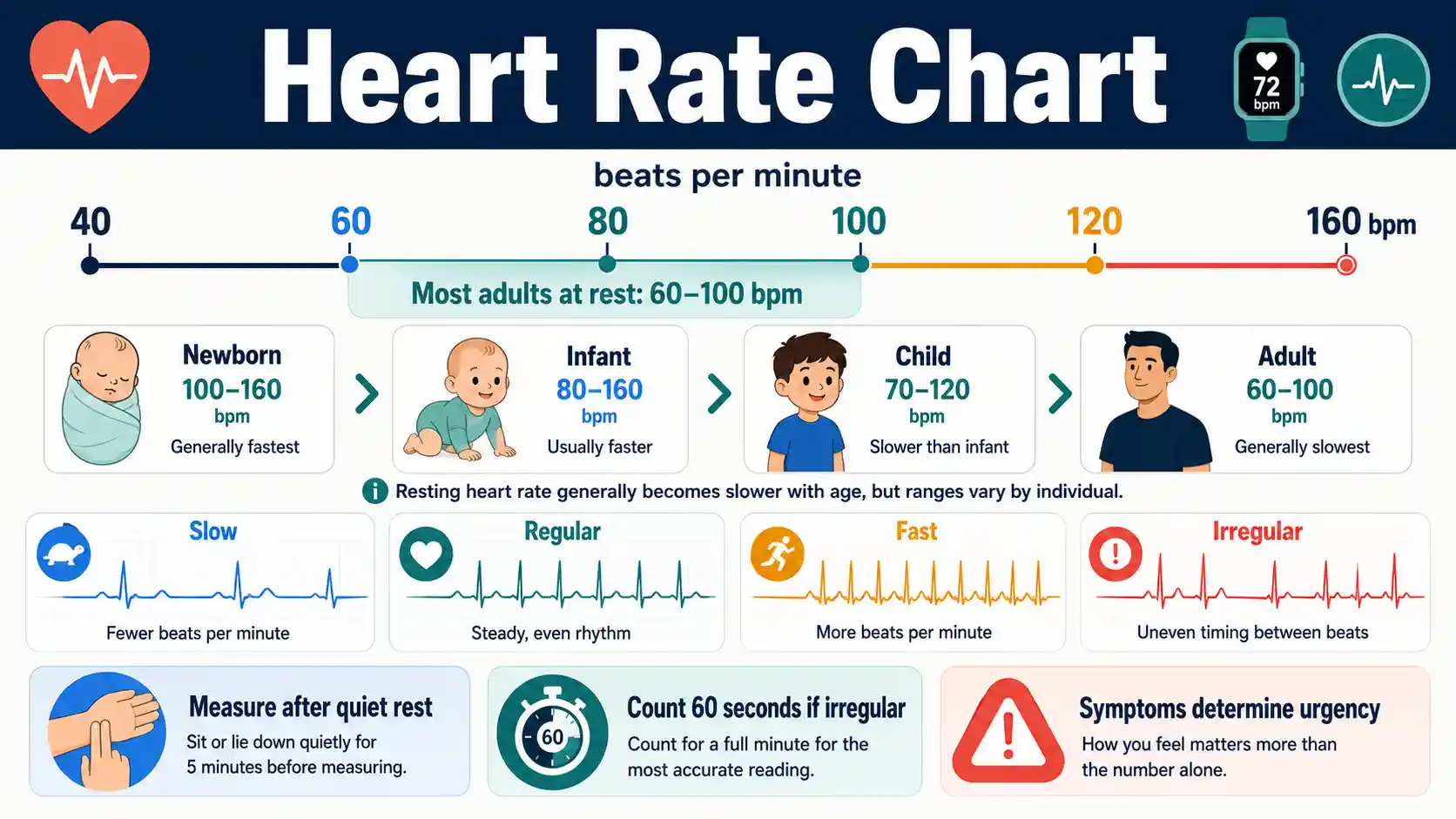
What is a normal heart rate?
For most adults who are calm, resting, and feeling well, 60 to 100 beats per minute is the commonly cited range. Infants and young children normally have faster resting rates, so age-specific references are required.
The American Heart Association defines heart rate as beats per minute and explains the usual adult resting range. The rhythm, symptoms, personal baseline, and measurement conditions matter as much as the number.
Most adults at rest
60–100 bpm
Use this range only when sitting or lying, calm, and feeling well.
Children
Use age-specific ranges
Normal resting pulse is generally faster in infants and young children.
Best manual count
60 seconds
Especially when the pulse is irregular, faint, very fast, very slow, or pediatric.
Emergency context
Symptoms change urgency
Chest pain, severe breathlessness, fainting, confusion, or collapse need emergency care.
Resting Heart Rate by Age Chart
These reference ranges apply to a resting pulse after adequate quiet rest. Children normally have faster resting rates than adults, and one reading must be interpreted with symptoms and circumstances.
Swipe horizontally inside the table to view every column.
| Age group | Reference resting range | Measurement context | Interpretation note |
|---|---|---|---|
| Newborn, 0–1 month | 70–190 bpm | Quiet or resting, not crying or feeding | Wide variation is expected; illness, temperature, sleep, and distress can change the rate |
| Infant, 1–11 months | 80–160 bpm | Resting and calm | Count for a full minute when the rhythm is difficult to feel or appears irregular |
| Child, 1–2 years | 80–130 bpm | Resting and settled | Recent activity, crying, fever, pain, and dehydration can raise the pulse |
| Child, 3–4 years | 80–120 bpm | Resting and calm | Compare repeated measurements taken under similar conditions |
| Child, 5–6 years | 75–115 bpm | Resting and calm | A single value outside the range is not a diagnosis |
| Child, 7–9 years | 70–110 bpm | Resting and calm | Symptoms and the child's usual baseline matter |
| Age 10 years and older, including adults | 60–100 bpm | Sitting or lying, calm, and feeling well | Persistent values outside this range should be discussed with a clinician |
| Well-trained athlete | Often 40–60 bpm | Resting and without concerning symptoms | A low athletic pulse can be normal, but fainting, weakness, chest pain, or breathlessness requires assessment |
bpm = heartbeats per minute.
- • MedlinePlus recommends resting for at least 10 minutes before measuring a resting pulse.
- • Ranges are broad screening references, not treatment thresholds. Sleep, fever, distress, medication, fitness, hydration, and illness can shift the rate.
- • For infants and children, measure when calm whenever possible and follow the advice of the child's clinician when a condition or medication changes the expected range.
Download or export
Use the correct age group and a true resting measurement
MedlinePlus lists broad resting pulse ranges by age and recommends at least 10 minutes of rest. Crying, feeding, fever, recent movement, pain, anxiety, dehydration, and sleep can shift a child or adult reading.
How to Measure Heart Rate and Pulse
A manual pulse is most useful when the person is at rest, the site is easy to feel, and the counting interval is long enough for the rhythm.
Swipe horizontally inside the table to view every column.
| Step or site | What to do | Useful for | Common mistake to avoid |
|---|---|---|---|
| Prepare | Rest quietly for at least 10 minutes before a resting measurement | Establishing a comparable baseline | Measuring immediately after walking, caffeine, stress, or exercise and calling it resting |
| Wrist, radial pulse | Place index and middle fingers on the thumb side of the inner wrist with light pressure | Most home measurements | Using the thumb, pressing too hard, or moving the fingers repeatedly |
| Neck, carotid pulse | Use two fingers on one side of the neck only and press gently | When the wrist pulse is difficult to find | Pressing both sides of the neck or applying strong pressure |
| Other pulse sites | Inside the elbow or top of the foot may be used when appropriate | Clinical examination or when common sites are unavailable | Assuming a difficult-to-find pulse is always harmless |
| Count 60 seconds | Count every beat for one full minute | Irregular, very slow, very fast, faint, or pediatric pulses | Using a short interval when beats are uneven |
| Count 30 seconds | Count for 30 seconds and multiply by 2 | A clearly regular adult pulse | Rounding or losing track of beats |
| Record context | Write down bpm, time, posture, symptoms, activity, and device or method | Finding trends and sharing useful information with a clinician | Recording only the number without circumstances |
| Repeat if unexpected | Sit quietly, check technique, and repeat after several minutes if safe to do so | Reducing one-off measurement error | Repeatedly checking during panic or delaying emergency care when severe symptoms are present |
- • The American Heart Association recommends counting the wrist pulse for 60 seconds for the most accurate manual reading.
- • Do not massage or strongly compress the neck. Seek professional help if a pulse is difficult to locate and the person is unwell.
- • A pulse gives rate and a sense of regularity. It does not replace an ECG when an arrhythmia is suspected.
Download or export
Adult Resting Heart Rate Interpretation Chart
For most adults who are calm, resting, and feeling well, 60–100 bpm is the commonly cited range. The number alone does not establish whether the rhythm is healthy.
Swipe horizontally inside the table to view every column.
| Resting finding | Common label | What it may mean | Appropriate next step |
|---|---|---|---|
| Below 60 bpm | Bradycardia by rate definition | Can be normal during sleep, in fit people, or with rate-slowing medication; can also occur with conduction or medical problems | Compare with usual baseline and symptoms; ask a clinician about persistent or unexplained readings |
| 60–100 bpm | Usual adult resting range | Often expected when sitting or lying, calm, and feeling well | Track trends rather than treating one number as a guarantee of health |
| Above 100 bpm | Tachycardia by rate definition | Can occur with exercise, fever, pain, stress, dehydration, stimulants, anemia, thyroid disease, infection, or arrhythmia | Rest and repeat if appropriate; seek clinical advice when persistent, unexplained, or symptomatic |
| Any rate with irregular rhythm | Possible arrhythmia or premature beats | May feel like fluttering, skipped beats, pauses, or pounding | Count for a full minute and arrange assessment when new, recurrent, or symptomatic |
| Sudden major change from personal baseline | Context-dependent abnormal change | May be more important than crossing a generic cutoff | Consider symptoms, medication changes, illness, and seek timely advice |
| Very high or very low with severe symptoms | Emergency warning pattern | May reduce effective blood flow or reflect a serious rhythm or medical problem | Call emergency services for chest pain, severe breathlessness, fainting, confusion, or collapse |
- • Bradycardia and tachycardia are rate descriptions, not complete diagnoses.
- • A regular pulse at 70 bpm can still coexist with heart disease, and an irregular pulse may need ECG confirmation even when the average rate is within 60–100 bpm.
- • People taking beta blockers or other rate-altering medicines should use the monitoring plan provided by their prescriber.
Download or export
Rate and rhythm
The same beats-per-minute value can hide different rhythms
A pulse count gives an average rate. The spacing between beats, how the episode started, how long it lasted, symptoms, and an ECG tracing help determine whether the rhythm is normal or abnormal.
Slow
<60Can be normal or clinically important
Typical adult resting range
60–100When calm, resting, and feeling well
Fast
>100Context and symptoms determine concern
Irregular
UnevenNeeds rhythm context, often ECG confirmation
Rate is not the same as rhythm
NHLBI describes bradycardia, tachycardia, premature beats, and other arrhythmias. A pulse count can suggest slow, fast, or uneven timing, but an ECG is needed to identify the electrical pattern.
Pulse count to beats-per-minute calculator
Enter the number of beats counted and the counting interval. For a calm resting measurement, the tool can compare the result with the selected broad age reference. It does not identify the rhythm or provide medical clearance.
Calculated pulse
72 bpm
Within the selected resting reference
The selected reference is 60–100 bpm. Being inside a broad range does not rule out an irregular rhythm or other health problem.
- Calculation
- 36 × 60 ÷ 30
- Selected reference
- 60–100 bpm
Limitation: This tool calculates an average rate. It cannot detect skipped beats, confirm an arrhythmia, judge circulation, or determine whether exercise is safe.
Factors That Change Heart Rate
Heart rate responds to the body's oxygen demand, temperature, nervous system, medicines, and illness. Interpretation improves when the measurement context is recorded.
Swipe horizontally inside the table to view every column.
| Factor | Typical direction | Why it changes heart rate | Interpretation tip |
|---|---|---|---|
| Exercise or physical work | Usually increases | Working muscles need more oxygenated blood | Use recovery and perceived effort context; do not compare directly with a quiet resting value |
| Sleep and deep relaxation | Often decreases | Metabolic demand and sympathetic drive fall | A low sleeping rate can be normal when there are no concerning symptoms |
| Fever or higher body temperature | Often increases | Metabolic demand rises and the body works to regulate temperature | Reassess after the illness improves and follow age-specific clinical advice |
| Stress, anxiety, excitement, or pain | Often increases | Adrenaline and sympathetic activity speed the heart | Rest, breathe normally, and repeat later when safe |
| Standing up | May briefly increase | The body adjusts circulation against gravity | A sustained rise with dizziness or fainting needs clinical assessment |
| Dehydration or blood loss | Often increases | The heart may compensate for reduced circulating volume | Seek care for significant dehydration, bleeding, weakness, or faintness |
| Fitness training | May lower resting rate | The heart may pump more blood per beat | Symptoms still matter; fitness does not make every low rate harmless |
| Beta blockers and some calcium-channel blockers | Often decrease | These medicines reduce rate or electrical conduction | Do not change a dose based on a chart; contact the prescriber for unexpected readings |
| Stimulants, nicotine, or some medicines | May increase | They can activate the cardiovascular and nervous systems | Review labels and medication history with a clinician |
| Illness or endocrine conditions | May increase or decrease | Infection, anemia, thyroid disorders, lung disease, and other conditions can alter rate | Persistent unexplained change deserves medical evaluation |
- • The same heart rate can have different meaning at rest, during exercise, during fever, or after a medication dose.
- • Body position usually causes only a small temporary change in healthy people. Symptoms and sustained changes are more important than a brief rise.
- • Do not use heart rate alone to judge hydration, infection severity, blood loss, or thyroid status.
Download or export
Heart Rate and Rhythm Pattern Chart
Rate describes how fast the heart beats. Rhythm describes timing and regularity. A pulse can be fast, slow, irregular, or any combination of these.
Swipe horizontally inside the table to view every column.
| Pattern | How it may feel or appear | Possible context | Why rate alone is insufficient |
|---|---|---|---|
| Regular and within usual range | Even spacing between beats | Normal sinus rhythm is possible | Only an ECG can define the electrical rhythm |
| Regular and fast | Rapid, evenly spaced beats | Exercise, fever, stress, dehydration, sinus tachycardia, or another tachycardia | The cause depends on onset, symptoms, duration, and ECG pattern |
| Regular and slow | Slow, evenly spaced beats | Sleep, fitness, medication, sinus-node or conduction problems | A rate below 60 can be normal or clinically important |
| Irregularly irregular | No repeating beat pattern | Atrial fibrillation is one possible cause | Pulse checks and wearables can flag irregularity but cannot confirm the diagnosis |
| Premature or extra beat pattern | A skipped sensation, pause, or stronger following beat | Premature atrial or ventricular beats, stimulants, stress, or other triggers | Frequency, origin, and risk require clinical context and sometimes monitoring |
| Sudden start and stop | Abrupt racing or pounding episode | Paroxysmal supraventricular tachycardia or another intermittent rhythm | An ECG during the episode or an ambulatory monitor may be needed |
| Pulse deficit or weak peripheral pulse | Some heartbeats may not be felt at the wrist | Irregular rhythm, low output, poor circulation, or measurement difficulty | The pulse count may differ from the actual heart rate measured by ECG |
- • Palpitations are a symptom description, not a rhythm diagnosis.
- • A normal-looking smartwatch rate does not exclude brief or intermittent arrhythmias.
- • New irregularity with chest pain, shortness of breath, fainting, or neurologic symptoms requires urgent evaluation.
Download or export
Heart Rate Monitoring Methods Chart
Different tools answer different questions. A rate reading is not the same as a diagnostic rhythm tracing.
Swipe horizontally inside the table to view every column.
| Method | What it measures | Best use | Main limitation |
|---|---|---|---|
| Manual pulse | Peripheral beats per minute and perceived regularity | Quick resting checks and symptom logs | Cannot identify the electrical rhythm or capture episodes that stop before measurement |
| Automatic blood-pressure monitor | Pulse rate during cuff measurement | Convenient combined blood pressure and pulse check | Motion and irregular rhythms can reduce reliability; device alerts are not diagnoses |
| Pulse oximeter | Pulse rate plus estimated oxygen saturation | Short-term spot checks when clinically appropriate | Cold fingers, poor circulation, motion, nail products, and signal quality can affect readings |
| Consumer watch or fitness tracker | Usually optical pulse trends; some devices also record a single-lead ECG | Activity, sleep, and personal trend awareness | Fit, skin contact, motion, algorithms, and device features vary; wellness data do not replace medical evaluation |
| 12-lead ECG or EKG | Electrical activity from multiple views for a short period | Most common clinical test for diagnosing an arrhythmia | May miss intermittent episodes that are not present during the recording |
| Holter monitor | Continuous ambulatory ECG, commonly for a day or longer | Frequent symptoms or rhythm assessment during normal activities | May miss rare events outside the recording window |
| Event or patch monitor | Longer ambulatory ECG, continuous or symptom-triggered depending on device | Less frequent intermittent symptoms | Requires correct wear and may not capture every event |
| Implantable loop recorder | Long-term rhythm monitoring under the skin | Very infrequent but concerning unexplained episodes | Requires a procedure and clinician-directed follow-up |
| Exercise stress test | ECG, rate, symptoms, and often blood pressure during controlled exertion | Exercise-related symptoms and cardiovascular response | Must be selected and interpreted for the individual clinical question |
- • NHLBI identifies the ECG as the most common test for diagnosing arrhythmias and describes ambulatory monitors for longer recording.
- • Consumer devices may be useful for trends or sharing a recording, but they should not be used to start, stop, or change treatment without professional advice.
- • Record the time and symptoms when an event occurs so a clinician can compare them with monitor data.
Download or export
A rate alert is different from a rhythm diagnosis
NHLBI identifies the ECG as the most common arrhythmia test and describes Holter, event, and loop monitors. Consumer devices can support trend awareness, but clinical decisions require the correct recording and interpretation.
Exercise Heart Rate Context Chart
Heart rate normally rises with activity, but a universal number does not describe the same effort for every person. This page avoids target-zone calculations because those require a separate, individualized discussion.
Swipe horizontally inside the table to view every column.
| Phase or population | Expected context | Useful observation | Important limitation |
|---|---|---|---|
| Before activity | Establish whether the person feels well and note the resting baseline | Unexpectedly high or low rate plus symptoms may be a reason to pause | One pre-exercise reading cannot determine exercise safety |
| Warm-up | Heart rate should rise gradually with increasing work | A smooth response usually matches increasing effort | Abrupt symptoms matter more than achieving a predicted number |
| Moderate relative intensity | Breathing and heart rate increase | CDC talk test: able to talk but not sing | The same bpm may be easy for one person and hard for another |
| Vigorous relative intensity | Marked increase in breathing and heart rate | Only a few words can be spoken before pausing for breath | Do not push through chest pain, faintness, or unusual breathlessness |
| Recovery | Rate falls after activity stops | Track how the rate and symptoms settle over time under similar conditions | Recovery depends on fitness, heat, hydration, medication, and workload |
| Beta blockers or rate-limiting medicine | Exercise heart rate may be lower than age-predicted formulas expect | Use clinician guidance and perceived effort | Do not alter medication to reach a target rate |
| Pregnancy | Heart rate and perceived effort can change across pregnancy | ACOG uses moderate-intensity symptoms and the talk test rather than one universal pulse target | Pregnancy complications and individual restrictions require obstetric guidance |
| Known heart or lung condition | Response may differ from population formulas | Use a prescribed plan, cardiac rehabilitation, or clinician-set limits when provided | Generic charts cannot establish a safe training ceiling |
- • CDC describes moderate relative intensity as about 5–6 on a 0–10 effort scale and vigorous intensity as beginning around 7–8.
- • Stop activity and seek urgent care for chest pain, fainting, severe shortness of breath, confusion, or a sustained dangerous-feeling rhythm.
- • A later ChartsLoom Heart Rate Zones Chart will explain common formulas and their limitations without turning estimates into medical clearance.
Download or export
Exercise intensity is individual
CDC explains relative intensity and the talk test: moderate activity usually allows talking but not singing, while vigorous effort allows only a few words before pausing. Medication, pregnancy, health conditions, heat, and fitness can make a generic heart-rate formula misleading.
Heart Rate Warning Signs and Next Steps
Symptoms, sudden change, persistence, and personal medical context are more important than one isolated number.
Swipe horizontally inside the table to view every column.
| Situation | Concern level | What to do | Why it matters |
|---|---|---|---|
| Sudden very high or very low rate with chest pain, severe shortness of breath, fainting, confusion, collapse, or signs of stroke | Emergency | Call local emergency services now | A dangerous rhythm or reduced blood flow may be present |
| Palpitations with fainting or near-fainting | Urgent or emergency depending on severity | Seek immediate medical assessment; do not drive yourself if unstable | Loss of consciousness can indicate inadequate blood flow or a serious rhythm |
| New irregular pulse with chest discomfort, breathlessness, weakness, or neurologic symptoms | Urgent | Obtain prompt medical evaluation | ECG testing may be needed to identify the rhythm and complications |
| Persistent resting rate above or below the person's normal range without severe symptoms | Contact clinician | Repeat under proper resting conditions and arrange advice | Medication, infection, dehydration, anemia, thyroid disease, or rhythm problems may contribute |
| Child with abnormal pulse plus poor feeding, lethargy, breathing difficulty, blue or pale color, or reduced responsiveness | Urgent or emergency | Seek pediatric emergency care based on severity | Infants and children can deteriorate quickly and have age-specific normal ranges |
| Rate alert from a wearable but the person feels well | Verify and monitor | Sit quietly, check a manual pulse when able, record context, and follow device or clinician guidance | Motion, contact, and algorithms can produce false alerts, but repeated findings deserve review |
| Brief isolated change after exercise, stress, or standing that settles and causes no symptoms | Usually observe | Record if recurrent and compare under similar conditions | Normal physiology can temporarily change the rate |
| Repeated skipped, fluttering, or pounding sensations | Clinical review | Keep a symptom and time log and discuss monitoring options | Intermittent rhythms may require an ECG, Holter, event, or patch monitor |
- • Emergency numbers and service names vary by country. Use the local emergency system where the person is located.
- • Do not delay emergency care to repeat measurements, charge a wearable, or search for a more reassuring number.
- • This chart does not determine whether a particular heart rate is safe for a person with a pacemaker, congenital heart disease, pregnancy complication, or prescribed rate target.
Download or export
Severe symptoms override the chart
Call local emergency services for a sudden very high or very low heart rate with chest pain, severe shortness of breath, fainting, confusion, collapse, or signs of stroke. Do not delay care to repeat the reading, charge a wearable, or search for a more reassuring number.
Heart rate chart FAQs
What is a normal resting heart rate for adults?
For most adults who are sitting or lying down, calm, and feeling well, 60 to 100 beats per minute is the commonly cited resting range. Fitness, sleep, temperature, medication, illness, and personal baseline can change what is expected.
Is a resting heart rate below 60 always dangerous?
No. A rate below 60 can occur during sleep, in well-trained people, or with rate-slowing medicine. It needs assessment when it is new, unexplained, persistent, or accompanied by weakness, dizziness, fainting, chest pain, or shortness of breath.
Is a resting heart rate above 100 always an arrhythmia?
No. Fever, pain, anxiety, dehydration, exercise, stimulants, infection, anemia, and other conditions can produce sinus tachycardia. A persistent or symptomatic resting rate above 100 should be evaluated to identify the cause.
Why is a child's heart rate faster than an adult's?
Infants and young children normally have faster resting pulses because their cardiovascular and metabolic needs differ from those of adults. Age-specific ranges should be used, and the child should be calm when possible.
How long should I rest before checking my resting pulse?
MedlinePlus advises resting for at least 10 minutes before a resting heart-rate measurement. Record whether you were sitting or lying, your symptoms, recent activity, and any factors such as fever, caffeine, or medication.
Should I count my pulse for 15, 30, or 60 seconds?
A full 60-second count is the safest general method and is especially important when the pulse is irregular, faint, very fast, very slow, or being measured in a child. A regular adult pulse may be counted for 30 seconds and doubled.
What is the difference between heart rate and pulse?
Heart rate is the number of heartbeats per minute. Pulse is the pressure wave felt in an artery with each effective heartbeat. They are often the same, but some irregular rhythms can create beats that are not felt strongly at the wrist.
Can a smartwatch diagnose an abnormal heart rhythm?
Some watches can flag rate changes or record a limited single-lead tracing, but features and accuracy vary. A consumer device does not replace a clinical ECG, medical history, examination, or professional interpretation.
What does an irregular pulse feel like?
People may describe fluttering, skipped beats, pauses, pounding, or an uneven rhythm. These sensations are called palpitations and do not identify the cause. New, recurrent, or symptomatic irregularity should be medically assessed.
Can fever raise heart rate?
Yes. A higher body temperature and the stress of illness commonly raise heart rate. The amount varies by age and condition, so the pulse should be interpreted alongside temperature, hydration, breathing, behavior, and other symptoms.
Does dehydration affect heart rate?
Dehydration can raise heart rate as the body tries to maintain circulation with less fluid volume. Significant thirst, reduced urination, weakness, dizziness, vomiting, diarrhea, or inability to drink may require medical assessment.
Why do beta blockers lower heart rate?
Beta blockers reduce the effect of stress hormones on the heart and are often prescribed to slow rate or lower workload. Do not change the dose to reach a chart value; follow the monitoring and action plan provided by the prescriber.
What heart rate is normal during exercise?
Exercise heart rate depends on age, fitness, medication, temperature, hydration, health conditions, and workload. Perceived effort and the talk test can provide useful context, while a generic formula cannot provide medical clearance or a precise safe limit.
When does a heart-rate change need emergency care?
Call local emergency services when a sudden very high or very low rate occurs with chest pain, severe shortness of breath, fainting, confusion, collapse, or signs of stroke. Do not delay care to repeat the reading.
What test confirms an arrhythmia?
An electrocardiogram, also called an ECG or EKG, is the most common diagnostic test. Intermittent symptoms may require a Holter monitor, patch or event monitor, stress test, or implantable loop recorder selected by a clinician.
Sources
URLs are shown as plain text for transparent reference. Table-specific source names link to this section.
American Heart Association — All About Heart Rate
Defines heart rate and pulse, gives the usual adult resting range, explains manual pulse measurement, lists factors that change heart rate, and identifies emergency symptoms.
https://www.heart.org/en/health-topics/high-blood-pressure/the-facts-about-high-blood-pressure/all-about-heart-rate-pulse
MedlinePlus, U.S. National Library of Medicine — Pulse
Provides resting pulse reference ranges from newborn through adulthood, preparation guidance, measurement methods, and reasons to discuss persistent or irregular readings with a clinician.
https://medlineplus.gov/ency/article/003399.htm
National Heart, Lung, and Blood Institute — Arrhythmias: Types
Explains bradycardia, tachycardia, premature beats, atrial and ventricular arrhythmias, and basic pulse counting.
https://www.nhlbi.nih.gov/health/arrhythmias/types
National Heart, Lung, and Blood Institute — Arrhythmias: Diagnosis
Describes electrocardiography, stress testing, Holter monitors, event monitors, and implantable loop recorders used to evaluate abnormal rhythms.
https://www.nhlbi.nih.gov/health/arrhythmias/diagnosis
MedlinePlus, U.S. National Library of Medicine — Arrhythmia
Summarizes common rhythm symptoms, possible contributing conditions, emergency symptoms, and the role of ECG and other tests.
https://medlineplus.gov/arrhythmia.html
Centers for Disease Control and Prevention — How to Measure Physical Activity Intensity
Explains relative exercise intensity and the talk test, emphasizing that the same activity can produce different effort levels in different people.
https://www.cdc.gov/physical-activity-basics/measuring/index.html
American College of Obstetricians and Gynecologists — Exercise During Pregnancy
Uses perceived effort and the talk test for moderate activity during pregnancy rather than a single universal heart-rate target.
https://www.acog.org/womens-health/faqs/exercise-during-pregnancy
U.S. Food and Drug Administration — General Wellness: Policy for Low Risk Devices
Distinguishes low-risk general wellness products from functions intended to diagnose, treat, or make clinical decisions.
https://www.fda.gov/regulatory-information/search-fda-guidance-documents/general-wellness-policy-low-risk-devices