Biology and neuroanatomy reference
Nervous System Chart
This nervous system chart explains the brain, spinal cord, cranial and spinal nerves, neurons, glial cells, autonomic divisions, and reflex pathways that coordinate sensation, movement, cognition, and organ function.
Neurological symptoms can arise from the brain, spinal cord, roots, plexuses, peripheral nerves, muscles, circulation, infection, medication, or metabolic conditions. A diagram cannot diagnose the source.
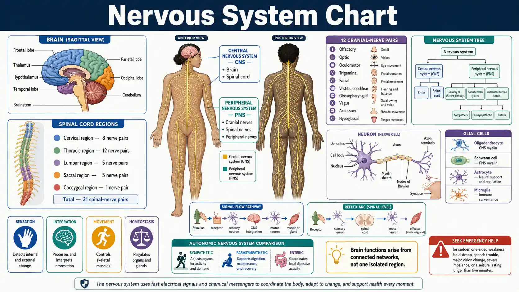
What are the main parts of the nervous system?
The central nervous system contains the brain and spinal cord. The peripheral nervous system contains cranial nerves, spinal nerves, ganglia, and sensory receptors that connect the CNS with skin, muscles, joints, organs, and glands. Functional pathways carry sensory input toward the CNS and motor output toward skeletal muscle or visceral effectors.
Central division
Brain and spinal cord
The CNS integrates sensory input, supports cognition, and coordinates motor and autonomic output.
Peripheral division
Nerves and ganglia
The PNS connects receptors, muscles, glands, and organs with the central nervous system.
Spinal nerves
31 pairs
Humans conventionally have 8 cervical, 12 thoracic, 5 lumbar, 5 sacral, and 1 coccygeal pair.
Cranial nerves
12 pairs
Cranial nerves support smell, vision, eye movement, facial sensation, hearing, balance, swallowing, voice, and other functions.
Essential nervous-system questions
These direct answers establish the definitions and limits needed to interpret the detailed charts correctly.
What does the nervous system do?
The nervous system detects changes, processes information, and coordinates movement, sensation, cognition, organ function, and homeostasis.
What is the central nervous system?
The central nervous system consists of the brain and spinal cord.
What is the peripheral nervous system?
The peripheral nervous system includes nerves and ganglia outside the brain and spinal cord that connect the CNS with the body.
What is a neuron?
A neuron is a specialized cell that receives, integrates, and transmits information through electrical and chemical signals.
What is a nerve?
A peripheral nerve is a bundled structure containing many axons, support cells, connective tissue, and blood vessels.
What is the difference between sensory and motor pathways?
Sensory pathways carry information toward the CNS, while motor pathways carry commands from the CNS to muscles and glands.
How many cranial nerves are there?
Humans have 12 conventionally numbered pairs of cranial nerves.
How many spinal nerves are there?
Humans have 31 pairs of spinal nerves.
What is the autonomic nervous system?
The autonomic nervous system regulates cardiac muscle, smooth muscle, glands, and many internal-organ functions.
What is a reflex arc?
A reflex arc links a receptor, sensory pathway, CNS integration center, motor pathway, and effector to produce a rapid patterned response.
Does one brain region control one function?
No. Most functions emerge from distributed networks that connect several cortical, subcortical, brainstem, cerebellar, and spinal regions.
Can a nervous-system chart diagnose symptoms?
No. Neurological diagnosis requires timing, examination, and often imaging, laboratory testing, or electrical studies.
Nervous System Divisions and Main Functions
The nervous system can be organized structurally as central versus peripheral and functionally as sensory, somatic motor, autonomic motor, and enteric pathways.
Swipe horizontally inside the table to view every column.
| Division | Main structures | Signal direction or role | Primary targets | Key distinction |
|---|---|---|---|---|
| Central nervous system (CNS) | Brain and spinal cord | Integrates information and coordinates responses | Neural circuits throughout the CNS | Contains central nuclei and tracts |
| Peripheral nervous system (PNS) | Cranial nerves, spinal nerves, ganglia, sensory receptors | Carries signals between the CNS and the body | Skin, muscles, joints, organs, glands | Contains peripheral nerves and ganglia |
| Sensory or afferent division | Somatic and visceral sensory neurons | Carries information toward the CNS — Toward the CNS | Receptors for touch, pain, temperature, stretch, chemicals, and special senses | Afferent means arriving at the CNS |
| Somatic motor division | Motor neurons to skeletal muscle | Carries motor commands away from the CNS — Away from the CNS | Skeletal muscle | Supports voluntary movement and somatic reflexes |
| Autonomic nervous system | Sympathetic and parasympathetic pathways | Regulates visceral motor output | Cardiac muscle, smooth muscle, glands — Visceral effectors | Usually operates without conscious control |
| Enteric nervous system | Neural networks in the gastrointestinal wall | Coordinates local digestive activity | Digestive smooth muscle, glands, blood vessels | Can function locally but communicates with the CNS and autonomic pathways |
| Sympathetic division | Thoracolumbar autonomic pathways and ganglia | Mobilizes resources and adjusts organs for demand | Heart, vessels, airways, glands, viscera | Effects vary by organ; not simply “on” |
| Parasympathetic division | Craniosacral autonomic pathways and terminal ganglia | Supports maintenance, digestion, and recovery functions | Eyes, glands, heart, lungs, digestive and pelvic organs | Effects vary by organ; not simply “off” |
Division names describe anatomical or functional organization. Real pathways often carry mixed sensory, motor, somatic, and autonomic fibers.
- • The brain and spinal cord form the CNS; nerves outside them are usually classified as PNS structures.
- • Somatic does not mean sensory only; the somatic system includes sensory input and motor output to skeletal muscle.
- • The enteric nervous system is extensive and locally active but remains connected to autonomic and central pathways.
Download or export
Signal-flow map
Nervous-system function follows linked input, integration, and output pathways
A useful nervous-system chart connects receptors, afferent neurons, CNS circuits, efferent neurons, and effectors. Real signals also receive continuous feedback from posture, organ state, attention, memory, and the environment.
Sensory input
Receptor → peripheral nerve → CNS
Sensory receptors convert physical or chemical changes into neural signals that travel toward the spinal cord and brain.
Integration
Spinal cord and brain networks
Interneurons and distributed circuits compare inputs, select responses, update memory, and coordinate reflexive and conscious processing.
Somatic output
CNS → motor neuron → skeletal muscle
Somatic motor neurons drive voluntary movement and participate in reflexes through direct connections with skeletal muscle fibers.
Autonomic output
CNS → ganglion → visceral effector
Autonomic pathways regulate cardiac muscle, smooth muscle, glands, and organ function through sympathetic and parasympathetic circuits.
Electrical signaling
Action potentials propagate along axons through controlled ion movement across the cell membrane.
Chemical signaling
Many synapses release neurotransmitters that excite, inhibit, or modulate the next cell.
Network behavior
Perception and movement emerge from connected circuits rather than one neuron or one brain region acting alone.
The nervous system is a network, not a simple wiring diagram
The OpenStax nervous-system overview organizes neural function as sensory input, integration, and motor output. Real circuits add parallel pathways, feedback, inhibition, memory, prediction, and continuous communication between central and peripheral structures.
Major Brain Regions and Representative Functions
Brain functions arise from distributed networks. The listed roles are useful orientation points, not exclusive one-region assignments.
Swipe horizontally inside the table to view every column.
| Region | Major components | Representative functions | Important connections | Interpretation limit |
|---|---|---|---|---|
| Cerebral cortex | Frontal, parietal, temporal, and occipital lobes plus insula | Perception, voluntary movement, language, planning, memory, attention | Thalamus, basal nuclei, cerebellum, brainstem, spinal cord | Functions are distributed across connected cortical networks |
| Frontal lobe | Prefrontal, premotor, motor, language-related regions | Planning, decision-making, voluntary movement, behavioral control, speech production in many people | Parietal cortex, basal nuclei, thalamus, cerebellum | Laterality and network organization vary |
| Parietal lobe | Somatosensory and association cortices | Touch and body-position processing, spatial attention, sensorimotor integration | Thalamus, frontal cortex, visual association areas | A sensory map is not a complete map of experience |
| Temporal lobe | Auditory cortex, medial temporal structures, association cortex | Hearing, language comprehension, memory, object and face processing | Limbic structures, frontal and parietal cortex | Memory and language rely on broader networks |
| Occipital lobe | Primary and association visual cortices | Early visual processing and interpretation | Thalamus, parietal and temporal visual streams | Vision also requires eyes, optic pathways, and association networks |
| Basal nuclei | Caudate, putamen, globus pallidus and related circuits | Movement selection, habit learning, motivation-related loops | Cortex, thalamus, brainstem | They modulate movement rather than directly powering muscles |
| Thalamus | Multiple relay and association nuclei | Routes and modulates most sensory information and participates in motor and cognitive loops — Major relay and modulation role | Cortex, basal nuclei, cerebellum, brainstem | Olfaction has a different initial relay pattern |
| Hypothalamus | Nuclei linked with pituitary and autonomic pathways | Homeostasis, endocrine control, temperature, hunger, thirst, circadian and autonomic regulation | Pituitary, brainstem, limbic system, cortex | It coordinates many systems rather than acting alone |
| Brainstem | Midbrain, pons, medulla | Conduction, cranial-nerve functions, arousal, breathing and cardiovascular control — Vital regulatory centers | Cerebrum, cerebellum, spinal cord, cranial nerves | Small lesions can affect several tightly packed pathways |
| Cerebellum | Cerebellar cortex, deep nuclei, peduncles | Coordination, timing, balance, motor learning, error correction — Coordination and motor learning | Cortex, brainstem, vestibular system, spinal pathways | It does not initiate voluntary movement by itself |
Regional functions are representative. Clinical localization uses symptom pattern, timing, examination, and imaging rather than one symptom-to-one-lobe matching.
- • The cerebral hemispheres contain gray-matter cortex over white matter and deeper nuclei.
- • The diencephalon includes the thalamus and hypothalamus among other structures.
- • The brainstem contains many cranial-nerve nuclei and long ascending and descending tracts.
Download or export
Spinal Cord Segments, Roots, and Nerve Counts
The spinal cord is organized into segments that give rise to 31 pairs of spinal nerves. Segment level and vertebral level are not identical in the lower spine.
Swipe horizontally inside the table to view every column.
| Region | Spinal nerve pairs | General distribution | Key structures | Important note |
|---|---|---|---|---|
| Cervical | 8 pairs: C1–C8 — Eight cervical nerve pairs | Neck, diaphragm, upper limbs, parts of trunk | Cervical enlargement; cervical plexus; brachial plexus contributions | There are eight cervical nerves but seven cervical vertebrae |
| Thoracic | 12 pairs: T1–T12 | Thoracic wall, much of abdominal wall, sympathetic outflow | Intercostal nerves and thoracic roots | Most thoracic anterior rami remain segmental rather than forming large limb plexuses |
| Lumbar | 5 pairs: L1–L5 | Lower abdominal wall and parts of lower limb | Lumbar enlargement; lumbar plexus contributions | Lower cord segments lie above similarly numbered vertebrae in adults |
| Sacral | 5 pairs: S1–S5 | Pelvis, posterior thigh, much of leg and foot, pelvic organs | Sacral plexus contributions and pelvic pathways | Sacral roots descend in the cauda equina before exiting |
| Coccygeal | 1 pair: Co1 | Small area near the coccyx | Coccygeal nerve | Distribution is limited and variable |
| Dorsal root | One per side at each segment | Carries sensory afferent fibers toward the spinal cord — Sensory input | Dorsal root ganglion contains sensory neuron cell bodies | Root injury can create segmental sensory symptoms |
| Ventral root | One per side at each segment | Carries motor efferent fibers away from the spinal cord — Motor output | Somatic motor and preganglionic autonomic axons where present | The mixed spinal nerve forms after dorsal and ventral roots join |
| Spinal nerve | 31 pairs total | Mixed sensory and motor distribution | Divides into dorsal and ventral rami | A peripheral nerve may contain fibers from several spinal roots |
Counts refer to paired spinal nerves: 8 cervical, 12 thoracic, 5 lumbar, 5 sacral, and 1 coccygeal pair.
- • The adult spinal cord usually ends above the lowest lumbar vertebrae; lower roots continue as the cauda equina.
- • Dermatomes are approximate skin territories linked mainly to one spinal root and overlap with neighboring roots.
- • A root, spinal nerve, plexus, and named peripheral nerve are different anatomical levels.
Download or export
Twelve Cranial Nerves and Core Functions
Cranial nerves carry sensory, motor, and autonomic fibers between the brain or brainstem and structures mainly in the head and neck, with important exceptions such as the vagus nerve.
Swipe horizontally inside the table to view every column.
| Number and name | Main type | Core functions | Representative test or clue | Important limit |
|---|---|---|---|---|
| I — Olfactory | Special sensory | Smell | Identify a familiar odor in each nostril when appropriate | Smell loss can arise from nasal or central causes |
| II — Optic | Special sensory | Vision and afferent pupillary light pathway | Visual acuity, fields, pupils, fundus | It is a CNS tract by development and myelination — Developmentally a CNS pathway |
| III — Oculomotor | Motor and parasympathetic | Most eye movements, eyelid elevation, pupil constriction | Eye position, movement, eyelid, pupil response | Pupil findings depend on lesion location and cause |
| IV — Trochlear | Motor | Superior oblique eye muscle | Downward gaze with the eye adducted | Subtle deficits may cause vertical or torsional diplopia |
| V — Trigeminal | Mixed | Facial sensation, corneal afferent limb, chewing | Facial sensation and jaw movement | Three major sensory divisions have different territories |
| VI — Abducens | Motor | Lateral rectus eye muscle | Abduction of the eye | Long intracranial course can make it vulnerable in several conditions |
| VII — Facial | Mixed and parasympathetic | Facial expression, taste anterior tongue, lacrimal and salivary functions | Facial movement, taste when indicated, corneal efferent limb | Central and peripheral facial weakness patterns differ |
| VIII — Vestibulocochlear | Special sensory | Hearing and balance | Hearing tests, nystagmus, vestibular assessment | Symptoms can originate in the ear, nerve, or central pathways |
| IX — Glossopharyngeal | Mixed and parasympathetic | Taste posterior tongue, pharyngeal sensation, parotid secretion | Swallowing and gag-related assessment when indicated | Functions overlap with vagal pathways |
| X — Vagus | Mixed and parasympathetic | Voice, swallowing, visceral sensation, thoracic and abdominal parasympathetic output — Broad visceral distribution | Voice, palate movement, swallowing | Extends well beyond the head and neck |
| XI — Accessory | Motor | Sternocleidomastoid and trapezius movement | Head turn and shoulder shrug against resistance | Weakness can reflect muscle or nerve injury |
| XII — Hypoglossal | Motor | Tongue movement | Tongue protrusion and articulation | Deviation patterns require full localization |
“Sensory,” “motor,” and “mixed” summarize major fiber classes. Several cranial nerves also carry parasympathetic fibers.
- • Cranial nerves I and II differ developmentally from typical peripheral nerves.
- • A bedside cranial-nerve examination is interpreted as a pattern, not as twelve isolated pass-or-fail tests.
- • Sudden diplopia, facial weakness, swallowing difficulty, or speech change requires clinical assessment and may be an emergency when abrupt.
Download or export
Major Nerve Plexuses and Representative Peripheral Nerves
Ventral rami from several spinal nerves combine in plexuses before redistributing fibers into named peripheral nerves. Root levels are commonly variable and overlapping.
Swipe horizontally inside the table to view every column.
| Plexus or pattern | Typical roots | Representative nerves | Main distribution | Clinical interpretation limit |
|---|---|---|---|---|
| Cervical plexus | C1–C4 | Phrenic nerve and cutaneous branches — Phrenic nerve supports breathing | Neck; diaphragm through the phrenic nerve | Root contributions overlap and may vary |
| Brachial plexus | C5–T1 | Musculocutaneous, axillary, radial, median, ulnar | Shoulder girdle and upper limb | One named nerve contains fibers from multiple roots |
| Thoracic segmental nerves | T1–T12 | Intercostal and subcostal nerves | Thoracic and abdominal walls | Most do not form a large limb plexus |
| Lumbar plexus | L1–L4 | Femoral, obturator, lateral femoral cutaneous | Anterior and medial thigh and parts of abdominal wall | Lumbar and sacral plexuses connect through the lumbosacral trunk |
| Sacral plexus | L4–S4 | Sciatic, superior and inferior gluteal, pudendal | Pelvis, posterior thigh, most of leg and foot | The sciatic nerve later divides mainly into tibial and common fibular components |
| Radial nerve | Mainly C5–T1 through brachial plexus | Posterior arm and forearm branches | Elbow, wrist, and finger extension; posterior limb sensation | Symptoms depend on the lesion level |
| Median nerve | Mainly C6–T1 through brachial plexus | Anterior forearm and hand branches | Many forearm flexors, thumb function, lateral palm digits | Carpal tunnel affects the nerve at the wrist, not every median-nerve fiber |
| Ulnar nerve | Mainly C8–T1 through brachial plexus | Forearm and hand branches | Many intrinsic hand muscles and medial hand sensation | Compression sites include elbow and wrist |
| Femoral nerve | L2–L4 through lumbar plexus | Motor and saphenous branches | Hip flexion contribution, knee extension, anterior thigh and medial leg sensation | Root and peripheral-nerve lesions produce different patterns |
| Sciatic nerve | L4–S3 through sacral plexus | Tibial and common fibular components | Posterior thigh and most motor and sensory pathways below the knee through branches | “Sciatica” describes symptoms, not one single diagnosis — Symptom label is not a diagnosis |
Root ranges are typical teaching patterns and can vary. Peripheral nerve territories overlap and should be correlated with examination findings.
- • Dorsal rami supply the back; ventral rami form most plexuses and supply the anterolateral trunk and limbs.
- • A dermatome maps a spinal root, while a peripheral sensory territory maps a named nerve.
- • Numbness or weakness cannot be localized reliably from one simplified map alone.
Download or export
Browser-only educational tool
Nervous System Structure Comparison Explorer
Select two structures or divisions to compare location, function, signal pattern, major components, and a practical localization clue.
Selection 1
Central nervous system
Division
Central
Location
Cranial cavity and vertebral canal
Main role
Integrates information and coordinates neural responses.
Signal pattern
Contains local circuits plus ascending, descending, and interregional pathways.
Key components
Brain and spinal cord
Localization clue
Central lesions can affect multiple functions below or opposite the lesion depending on pathway.
Selection 2
Peripheral nervous system
Division
Peripheral
Location
Nerves and ganglia outside the brain and spinal cord
Main role
Connects sensory receptors and effectors with the CNS.
Signal pattern
Carries afferent sensory and efferent motor fibers.
Key components
Cranial nerves, spinal nerves, ganglia, receptors
Localization clue
Peripheral injury often follows a root, plexus, or named-nerve pattern.
Neuron Parts and Major Glial Cell Functions
Neurons transmit and process signals, while glial cells support, insulate, protect, nourish, and regulate neural environments. Both are essential to nervous-system function.
Swipe horizontally inside the table to view every column.
| Cell or structure | Location | Main role | Key feature | Common misconception |
|---|---|---|---|---|
| Dendrites | Neuron input region | Receive synaptic and sensory signals | Branching processes with receptors | They are not always passive cables |
| Cell body or soma | CNS nuclei, cortex, ganglia, peripheral receptors | Maintains the cell and integrates many inputs | Contains nucleus and organelles | The soma is not the whole neuron |
| Axon | Extends from neuron to targets | Conducts action potentials toward terminals | May be very short or very long | A nerve is a bundle of axons, not one axon |
| Myelin sheath | Around selected CNS and PNS axons | Increases conduction efficiency and supports axons | Interrupted by nodes of Ranvier | Not every axon is myelinated |
| Synaptic terminal | End of an axon branch | Releases neurotransmitter or communicates electrically | Forms specialized contact with target cell | Synapses do not always excite the next cell |
| Astrocytes | CNS | Support extracellular balance, synapses, metabolism, and blood–brain barrier relationships | Highly branched glial cells | They do more than structural support |
| Oligodendrocytes | CNS | Myelinate segments of multiple CNS axons — CNS myelin | One cell can support several internodes | They are not the PNS myelinating cell |
| Schwann cells | PNS | Myelinate one segment of one peripheral axon or support unmyelinated axons — PNS myelin | Wrap peripheral axons | One Schwann cell does not myelinate many separate internodes |
| Microglia | CNS | Immune surveillance and response to injury or debris — Immune surveillance | Resident immune-related cells | They are not neurons |
| Ependymal cells | CNS ventricles and central canal | Line fluid-filled spaces and participate in cerebrospinal-fluid interfaces | Ciliated or specialized epithelial-like lining | They do not form peripheral myelin |
| Satellite cells | PNS ganglia | Support neuron cell bodies and regulate local environment | Surround somas in ganglia | They are different from skeletal-muscle satellite cells |
Cell roles are simplified. Neural signaling depends on ion channels, membrane potentials, synapses, metabolism, blood flow, and network activity.
- • An action potential is a regenerative electrical change in membrane voltage, not electricity flowing through an empty wire.
- • Myelin changes conduction properties but does not create the underlying neural message.
- • Glial dysfunction can alter neural function even when neurons remain present.
Download or export
Neurons and glia form one functional tissue system
The OpenStax nervous-tissue guide describes neurons alongside astrocytes, oligodendrocytes, Schwann cells, microglia, ependymal cells, and satellite cells. Glia regulate the environment in which neural signaling occurs and are not inert packing material.
Sympathetic, Parasympathetic, and Enteric Effects
Autonomic divisions often produce contrasting effects, but organ responses are receptor-specific and coordinated rather than a universal on-versus-off switch.
Swipe horizontally inside the table to view every column.
| Target or function | Sympathetic tendency | Parasympathetic tendency | Enteric or local role | Interpretation note |
|---|---|---|---|---|
| Heart | Usually increases rate and contractile force | Usually slows sinoatrial rate | No primary independent cardiac network | Baseline autonomic tone and reflexes matter |
| Pupil | Dilates through radial iris muscle | Constricts and supports near focus | Not applicable | Light reflex also requires sensory and brainstem pathways |
| Airways | Promotes bronchodilation | Can promote bronchoconstriction and secretions | Not applicable | Respiratory control also depends on brainstem, hormones, and local signals |
| Salivary glands | Produces context-dependent secretion, often more viscous | Promotes watery secretion | Not applicable | Both divisions can stimulate secretion |
| Stomach and intestines | Generally reduces motility and secretion during acute demand | Generally increases digestive activity | Coordinates local motility, secretion, and reflexes — Local enteric coordination | Digestive control is distributed |
| Bladder | Supports storage through detrusor relaxation and outlet effects | Supports voiding through detrusor contraction | Local sensory and reflex circuits contribute | Voluntary and somatic pathways also regulate urination |
| Blood vessels | Major direct control of vascular smooth muscle in many beds | Limited direct supply to most vessels | Local metabolic control is important | Effects depend on receptor type and tissue |
| Sweat glands | Stimulates sweating through sympathetic pathways | No major direct role | Not applicable | Sympathetic fibers to sweat glands commonly use acetylcholine — Important neurotransmitter exception |
| Sexual function | Contributes to ejaculation and some vascular responses | Contributes to erection and glandular responses | Pelvic local circuits contribute | Both divisions and somatic pathways cooperate |
Effects are common tendencies in healthy physiology. Drugs, disease, receptor subtype, organ state, and reflex context can change the response.
- • Sympathetic and parasympathetic divisions can be active at the same time and may cooperate rather than oppose each other.
- • Autonomic pathways usually use a two-neuron motor chain from CNS to target, with a ganglion between neurons.
- • The autonomic nervous system controls cardiac muscle, smooth muscle, and glands; skeletal muscle uses somatic motor neurons.
Download or export
Reflex Arc Components and Example Pathways
A reflex is a rapid, patterned response to a stimulus. Many reflexes are integrated in the spinal cord or brainstem while information also travels to higher centers.
Swipe horizontally inside the table to view every column.
| Reflex component or example | Input | Integration | Output | Key teaching point |
|---|---|---|---|---|
| Receptor | Detects stretch, pain, pressure, light, chemicals, or another stimulus | Converts stimulus into neural activity | Sensory neuron is activated | The receptor determines stimulus sensitivity |
| Sensory neuron | Carries afferent signal | Enters spinal cord or brainstem | Activates interneurons or motor neurons | Cell body often lies in a sensory ganglion |
| Integration center | Receives sensory input | One or more synapses in CNS gray matter | Selects patterned motor response | Some reflexes are monosynaptic; many are polysynaptic |
| Motor neuron | Receives CNS output | Cell body in CNS motor region | Carries efferent signal to effector | Somatic motor neurons directly innervate skeletal muscle |
| Effector | Receives motor signal | Muscle or gland responds | Produces movement or secretion | Autonomic reflexes use visceral effectors |
| Patellar stretch reflex | Quadriceps muscle spindle stretch | Spinal monosynaptic excitation plus inhibitory interneuron pathway — Classic monosynaptic component | Quadriceps contracts; hamstring activity is reduced | Tests an arc involving peripheral nerve, roots, spinal cord, and muscle |
| Withdrawal reflex | Noxious skin stimulus | Polysynaptic spinal circuits | Flexors activate and extensors are inhibited | Protective response begins before conscious pain interpretation — Spinal response precedes awareness |
| Crossed-extensor reflex | Strong withdrawal input in one limb | Spinal interneuron network crosses midline | Opposite limb extends to support weight | Coordinates both sides of the body |
| Pupillary light reflex | Retinal light input | Midbrain bilateral relay | Both pupils constrict through parasympathetic output | Direct and consensual responses test linked pathways |
| Autonomic baroreflex | Stretch receptors sense pressure-related vessel-wall change | Brainstem cardiovascular centers | Heart and vascular autonomic output adjusts | Maintains short-term blood-pressure stability |
Reflex diagrams show the minimum teaching pathway. Real responses include modulation from the brain, attention, posture, medications, fatigue, and disease.
- • A normal reflex requires an intact receptor, sensory nerve, CNS synapse, motor pathway, neuromuscular junction, and muscle or gland.
- • A reflex can be absent, reduced, normal, brisk, or asymmetric; interpretation depends on the full neurological examination.
- • Do not intentionally test painful or dangerous reflexes outside appropriate clinical training.
Download or export
Neural maps simplify overlap, laterality, and anatomical variation
Dermatomes overlap, peripheral nerves contain fibers from several roots, brain functions use distributed networks, and autonomic effects depend on receptors and organ state. Symptoms also change with timing, vascular territory, compensation, and individual anatomy.
Maps are approximate
Dermatomes, nerve territories, and cortical maps overlap and vary among people.
One symptom has many causes
Weakness, numbness, dizziness, pain, and tremor can arise from neural or non-neural systems.
Timing changes urgency
Sudden focal deficits demand emergency assessment, while persistent or progressive symptoms still need clinical review.
Neurologic Warning Signs and Appropriate Action
Sudden or rapidly progressive neurological symptoms can reflect time-sensitive brain, spinal cord, nerve, or systemic disorders. This table supports escalation, not diagnosis.
Swipe horizontally inside the table to view every column.
| Pattern | Examples | Why it matters | Recommended action | Do not |
|---|---|---|---|---|
| Possible stroke or TIA | Sudden one-sided weakness or numbness, facial droop, speech difficulty, vision loss, severe imbalance, or sudden severe headache | Brain blood flow may be interrupted or bleeding may be present | Call emergency services immediately, even if symptoms improve — Emergency action | Do not drive yourself or wait for symptoms to pass |
| Prolonged or repeated seizure | Seizure lasts more than 5 minutes, repeats without recovery, occurs in water, causes injury, or breathing remains difficult | Prolonged seizure and impaired breathing can be life-threatening | Call emergency services and provide seizure first aid — Seizure emergency criteria | Do not restrain the person or put objects in the mouth |
| Sudden paralysis or rapidly worsening weakness | New inability to move a limb, spreading weakness, or weakness with breathing or swallowing difficulty | Can involve brain, spinal cord, peripheral nerves, muscles, or metabolic causes | Seek emergency assessment | Do not assume it is fatigue or a pinched nerve |
| New loss of consciousness or severe confusion | Collapse, inability to awaken normally, marked disorientation, or abrupt behavioral change | May reflect seizure, stroke, injury, infection, intoxication, or metabolic disturbance | Call emergency services | Do not give food, drink, or medication to an unresponsive person |
| Head injury with danger signs | Worsening headache, repeated vomiting, seizure, unequal pupils, increasing drowsiness, weakness, or confusion after trauma | May indicate significant brain injury or bleeding | Seek emergency care | Do not return to sport or risky activity |
| Fever with stiff neck or altered mental state | Fever plus severe headache, neck stiffness, confusion, unusual sleepiness, or seizure | Serious infection or inflammation may affect the nervous system | Seek urgent emergency assessment | Do not delay because a rash is absent |
| Progressive numbness or weakness | Symptoms spread over hours or days, affect both sides, or impair walking, hand use, breathing, or bladder function | Progressive nerve or spinal-cord disorders can worsen rapidly | Obtain urgent medical assessment | Do not rely only on a symptom map |
| New bowel or bladder dysfunction with saddle numbness | Urinary retention or incontinence with numbness around the groin or new leg weakness | May signal compression of lower spinal nerve roots | Seek emergency assessment — Possible cauda equina emergency | Do not postpone care for routine back-pain treatment |
| Persistent focal symptoms | Ongoing numbness, weakness, tremor, balance trouble, vision change, or recurrent headaches | Non-emergency neurological disorders still require evaluation | Arrange prompt clinical assessment | Do not self-diagnose from one chart |
Emergency systems and phone numbers differ by country. Use the local emergency number for sudden severe neurological symptoms.
- • Stroke symptoms can improve temporarily during a TIA, but this remains a medical emergency.
- • During a convulsive seizure, protect the person from injury, turn them onto one side when possible, and time the event.
- • This page does not replace examination, glucose testing, imaging, laboratory tests, or emergency triage.
Download or export
Sudden focal neurological symptoms require emergency action
The CDC stroke warning guide lists sudden one-sided weakness or numbness, speech trouble, vision change, loss of balance, and a sudden severe headache among warning signs. Call the local emergency number immediately, even when symptoms improve.
Frequently asked questions
What are the two main structural divisions of the nervous system?
The central nervous system contains the brain and spinal cord. The peripheral nervous system contains the nerves and ganglia that connect the CNS with the rest of the body.
What is the difference between afferent and efferent nerves?
Afferent pathways carry sensory information toward the central nervous system. Efferent pathways carry motor commands away from the central nervous system.
How many pairs of cranial nerves are there?
Humans have 12 conventionally numbered pairs of cranial nerves. Their functions include smell, vision, eye movement, facial sensation, hearing, balance, swallowing, voice, and tongue movement.
How many pairs of spinal nerves are there?
Humans have 31 pairs of spinal nerves: 8 cervical, 12 thoracic, 5 lumbar, 5 sacral, and 1 coccygeal pair.
Why are there eight cervical nerves but seven cervical vertebrae?
Cervical nerves C1 through C7 exit above their matching vertebrae, C8 exits between C7 and T1, and lower spinal nerves then exit below the similarly numbered vertebra.
What is the difference between a nerve and a neuron?
A neuron is one specialized cell. A peripheral nerve is a bundled structure containing many axons plus connective tissue, blood vessels, and support cells.
What do glial cells do?
Glial cells support neural metabolism, myelination, immune surveillance, extracellular balance, synaptic function, and fluid interfaces. They are active participants in nervous-system function.
What is myelin?
Myelin is a multilayered membrane sheath around selected axons that improves conduction efficiency and supports axonal function. Oligodendrocytes form CNS myelin, while Schwann cells form PNS myelin.
What is a ganglion?
A ganglion is a cluster of neuron cell bodies in the peripheral nervous system. A cluster of neuron cell bodies inside the CNS is generally called a nucleus.
What is a tract?
A tract is a bundle of axons inside the central nervous system. A comparable bundle in the peripheral nervous system is generally called a nerve.
Is the sympathetic nervous system always harmful or stressful?
No. Sympathetic activity is normal and essential for blood-pressure control, temperature regulation, exercise, alertness, and organ adjustment. Problems arise when regulation is impaired, not because the system exists.
Does the parasympathetic system simply switch the sympathetic system off?
No. The two divisions have organ-specific effects and can cooperate, oppose each other, or act at different times. Many tissues also use local, hormonal, and enteric regulation.
Does a reflex happen without the brain?
Some reflex responses are integrated in the spinal cord or brainstem before conscious awareness, but the brain still receives information and can modulate many reflexes.
Can a dermatome chart diagnose a pinched nerve?
No. Dermatomes overlap and real symptoms may arise from a spinal root, plexus, peripheral nerve, spinal cord, brain, muscle, joint, or systemic condition.
When is sudden numbness or weakness an emergency?
Sudden numbness or weakness, especially on one side or with facial droop, speech trouble, vision change, or severe imbalance, requires immediate emergency assessment for possible stroke.
When should emergency services be called for a seizure?
Call emergency services when a seizure lasts longer than five minutes, repeats without recovery, causes serious injury, occurs in water, or is followed by breathing difficulty or failure to awaken normally.
Sources
These anatomy, physiology, peripheral-nerve, neurological-examination, stroke, and seizure references support the divisions, functions, counts, pathways, and warning guidance used throughout the page.
OpenStax — Basic Structure and Function of the Nervous System
https://openstax.org/books/anatomy-and-physiology-2e/pages/12-1-basic-structure-and-function-of-the-nervous-system
Defines the central and peripheral nervous systems, sensory input, integration, motor output, gray matter, white matter, nuclei, ganglia, tracts, and nerves.
OpenStax — Nervous Tissue
https://openstax.org/books/anatomy-and-physiology-2e/pages/12-2-nervous-tissue
Describes neurons, dendrites, axons, myelin, synapses, action potentials, and major glial-cell types in the CNS and PNS.
OpenStax — The Central Nervous System
https://openstax.org/books/anatomy-and-physiology-2e/pages/13-2-the-central-nervous-system
Reviews the cerebrum, diencephalon, brainstem, cerebellum, spinal cord, gray matter, white matter, and major functional regions.
OpenStax — The Peripheral Nervous System
https://openstax.org/books/anatomy-and-physiology-2e/pages/13-4-the-peripheral-nervous-system
Covers cranial nerves, spinal nerves, plexuses, sensory and motor pathways, ganglia, and peripheral nerve organization.
OpenStax — Divisions of the Autonomic Nervous System
https://openstax.org/books/anatomy-and-physiology-2e/pages/15-1-divisions-of-the-autonomic-nervous-system
Compares sympathetic and parasympathetic pathways, autonomic ganglia, neurotransmitters, and visceral effectors.
OpenStax — Overview of the Neurological Exam
https://openstax.org/books/anatomy-and-physiology-2e/pages/16-1-overview-of-the-neurological-exam
Explains how mental status, cranial nerves, sensation, motor function, coordination, gait, and reflexes are assessed together.
NCBI Bookshelf — StatPearls — Neuroanatomy, Spinal Cord
https://www.ncbi.nlm.nih.gov/books/NBK559056/
Describes spinal-cord organization, ascending and descending pathways, 31 pairs of spinal nerves, and segmental anatomy.
NCBI Bookshelf — StatPearls — Neuroanatomy, Sensory Nerves
https://www.ncbi.nlm.nih.gov/books/NBK539846/
Reviews sensory receptors, afferent pathways, somatosensory processing, dermatomes, and peripheral sensory organization.
National Institute of Neurological Disorders and Stroke — Peripheral Neuropathy
https://www.ninds.nih.gov/health-information/disorders/peripheral-neuropathy
Defines the peripheral nervous system as the communication network linking the brain and spinal cord with the rest of the body and describes neuropathy symptoms.
National Institute of Neurological Disorders and Stroke — Glossary of Neurological Terms
https://www.ninds.nih.gov/health-information/disorders/glossary-neurological-terms
Provides public definitions for neurological structures, symptoms, peripheral nerves, and common clinical terminology.
Centers for Disease Control and Prevention — Signs and Symptoms of Stroke
https://www.cdc.gov/stroke/signs-symptoms/index.html
Lists sudden one-sided weakness or numbness, speech difficulty, vision trouble, balance loss, and severe headache as emergency stroke warning signs.
Centers for Disease Control and Prevention — First Aid for Seizures
https://www.cdc.gov/epilepsy/first-aid-for-seizures/index.html
Explains seizure first aid and when to call emergency services, including seizures lasting longer than five minutes or repeated seizures without recovery.